Review
. 2008 Jul-Aug;14(7-8):502-16.
doi: 10.2119/2008-00006.Legrand.

Renal hypoxia and dysoxia after reperfusion of the ischemic kidney

Affiliations
Review

Renal hypoxia and dysoxia after reperfusion of the ischemic kidney

Matthieu Legrand et al. Mol Med. 2008 Jul-Aug.

Abstract

Ischemia is the most common cause of acute renal failure. Ischemic-induced renal tissue hypoxia is thought to be a major component in the development of acute renal failure in promoting the initial tubular damage. Renal oxygenation originates from a balance between oxygen supply and consumption. Recent investigations have provided new insights into alterations in oxygenation pathways in the ischemic kidney. These findings have identified a central role of microvascular dysfunction related to an imbalance between vasoconstrictors and vasodilators, endothelial damage and endothelium-leukocyte interactions, leading to decreased renal oxygen supply. Reduced microcirculatory oxygen supply may be associated with altered cellular oxygen consumption (dysoxia), because of mitochondrial dysfunction and activity of alternative oxygen-consuming pathways. Alterations in oxygen utilization and/or supply might therefore contribute to the occurrence of organ dysfunction. This view places oxygen pathways' alterations as a potential central player in the pathogenesis of acute kidney injury. Both in regulation of oxygen supply and consumption, nitric oxide seems to play a pivotal role. Furthermore, recent studies suggest that, following acute ischemic renal injury, persistent tissue hypoxia contributes to the development of chronic renal dysfunction. Adaptative mechanisms to renal hypoxia may be ineffective in more severe cases and lead to the development of chronic renal failure following ischemia-reperfusion. This paper is aimed at reviewing the current insights into oxygen transport pathways, from oxygen supply to oxygen consumption in the kidney and from the adaptation mechanisms to renal hypoxia. Their role in the development of ischemia-induced renal damage and ischemic acute renal failure are discussed.


Figures
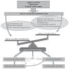
Figure 1
Figure 1
Synthetic representation of mechanisms involved in renal tissue hypoxia leading to acute and chronic renal failure after ischemia-reperfusion. Renal tissue hypoxia reflects an imbalance between oxygen supply and oxygen consumption. Renal tissue hypoxia participates to further tubular damage and acute renal failure. Recently the role of renal hypoxia has been proposed in development of chronic renal failure. HIF, hypoxia-inducible factor; –, protect; +, promote.
Figure 2
Figure 2
Interaction between microvascular injury and tubular damage before (A) and after (B) renal ischemia-reperfusion. PMN, polymorphonuclear.
Figure 3
Figure 3
Influence of NO and O2 on mitochondria activity. The mitochondrial electron transport chain includes four multiple subunit enzyme complexes of proteins (complexes I–IV), ATP synthase (complex V), and the adenine nucleotide translocator (ANT). Electrons are collected at the complexes I (NADH dehydrogenase) and II (succinate dehydrogenase) after oxidation of nicotinamide adenine dinucleotide (NADH) and of succinate, respectively, is transferred to coenzyme Q (CoQ) to give reduced CoQ. Electrons from reduced CoQ are transferred first to complex III (cytochrome bc1), then to cytochrome c (cyt c) and followed by complex IV (cytochrome c oxidase, COX), and finally to oxygen (O2) to give water (H2O). An electrochemical gradient is generated between the mitochondrial matrix and the intermembrane space by pumping protons through the inner membrane using the energy generated by the electron flow. This electrochemical gradient is then used to reintroduce protons through the complex V (ATP synthase) and to convert inorganic phosphate and ADP to ATP. The ATP, generated in mitochondria, is then exchanged for cytosolic ADP by the ANT. With increase in NO/O2 ratio, NO competitively inhibits complex IV associated with an electron leakage of the complexes chain and finally favors the generation of superoxide anions (O2−•). Hydrogen peroxide (H2O2) generated from O2−• by superoxide dismutase (SOD) contributes to hypoxia adaptation response via stabilization of HIF-α. If prolonged production of O2−• overwhelms capacity of the mitochondrial’s antioxidant system including the superoxide dismutase (MnSOD) and glutathione peroxidase (GSHpx), formation of large amount of peroxynitrite (ONOO–) and H2O2, precursor of the hydroxyl radical (OH•), via the Fenton reaction occurs. Peroxynitrite further inhibits the mitochondrial Mn-superoxide dismutase by nitration. Cytosolic (xanthine oxydase and cytochrome P450 reductases, and NOS, myeloperoxidase) and membrane (NADPH oxidase, the cytochrome P450) intramitochondrial ROS overproduction leads to cell necrosis or apoptosis.

Similar articles

Cited by

References

    1. Brady HR, Singer GG. Acute renal failure. Lancet. 1995;346:1533–40. - PubMed
    1. Bagshaw SM. The long-term outcome after acute renal failure. Curr Opin Crit Care. 2006;12:561–6. - PubMed
    1. Korkeila M, Ruokonen E, Takala J. Costs of care, long-term prognosis and quality of life in patients requiring renal replacement therapy during intensive care. Intensive Care Med. 2000;26:1824–31. - PubMed
    1. Manns B, et al. Cost of acute renal failure requiring dialysis in the intensive care unit: clinical and resource implications of renal recovery. Crit Care Med. 2003;31:449–55. - PubMed
    1. Bell M, Martling CR. Long-term outcome after intensive care: can we protect the kidney? Crit Care. 2007;11:147. - PMC - PubMed