Review
. 2008 Aug;237(8):1982-92.
doi: 10.1002/dvdy.21554.

Intraflagellar transport and the sensory outer segment of vertebrate photoreceptors

Affiliations
Review

Intraflagellar transport and the sensory outer segment of vertebrate photoreceptors

Christine Insinna et al. Dev Dyn. 2008 Aug.

Abstract

Analysis of the other segments of rod and cone photoreceptors in vertebrates has provided a rich molecular understanding of how light absorbed by a visual pigment can result in changes in membrane polarity that regulate neurotransmitter release. These events are carried out by a large group of phototransduction proteins that are enriched in the outer segment. However, the mechanisms by which phototransduction proteins are sequestered in the outer segment are not well defined. Insight into those mechanisms has recently emerged from the findings that outer segments arise from the plasma membrane of a sensory cilium, and that intraflagellar transport (IFT), which is necessary for assembly of many types of cilia and flagella, plays a crucial role. Here we review the general features of outer segment assembly that may be common to most sensory cilia as well those that may be unique to the outer segment. Those features illustrate how further analysis of photoreceptor IFT may provide insight into both IFT cargo and the role of alternative IFT kinesins.


Figures

Figure 1
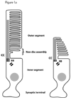
Figure 1. The photoreceptor sensory OS
A. Schematic illustrating the structure of rod and cone photoreceptors. The sensory organelle is composed of a stack of discs that initiated from the ciliary membrane at the base of the cilium. The dendritic portion extending from the basal body (BB) to the nucleus is called the inner segment. The cell body includes the nucleus and a short axon that terminates into a synapse. B. Simplified diagram of the phototransduction cascade taking place in the OS in the dark or upon light activation. This version does not include events involved in the deactivation of rhodopsin.
Figure 2
Figure 2. The photoreceptor cilium has a distinct transition zone and distal domain enriched in rhodopsin prior to the formation of discs
Rhodopsin labeling indicated by ferritin is enriched in the distal end and is much less abundant in the transition zone (between the arrows). Scale bar: 0.5 µm. Image was published previously (Besharse, 1986) and is reproduced here with permission of the publisher.
Figure 3
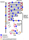
Figure 3. Simplified diagram illustrating the sorting problem for OS membrane proteins during disc formation
Membrane proteins are transported into vesicles from the Golgi complex to the base of the cilium. Once in the OS, they segregate to distinct compartments of the discs or the plasma membrane.
Figure 4
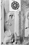
Figure 4. The structure of the connecting cilium is that of a transition zone
Images of a freeze fracture replica (A) and a conventionally stained EM section (B) oriented with the OS above and the inner segment below. Arrows indicate ciliary necklaces in A and bead-like membrane structures in B. Note the presence of fibrils in the extracellular space extending from the cilium membrane. The inset is a cross section showing doublet microtubules and the Y-shaped microtubule-membrane cross-linkers. Magnifcation bars are 0.1 µm for all three images. Image was published previously (Besharse, 1986) and is reproduced here with permission of the publisher.
Figure 5
Figure 5. Diagram showing similarities between C.elegans amphid channel cilia (left) and the photoreceptor cilium (right)
Both sensory cilia present a bipartite structure with a proximal segment composed of nine microtubule doublets with singlets towards the distal end.
Figure 6
Figure 6. Failure of OS formation in zebrafish embryos depleted of Kif17 protein
A. The retina of a 3 days old wild type embryo develops normal OS (arrowhead). B. Embryos injected with an antisense morpholino directed against Kif17 mRNA fail to develop OS (arrowhead) and present a small degree of disorganization of the retina layers. Scale bar in A 10 µm. C. EM picture of wild type photoreceptors with normal OS (arrowhead). Scale bar: 3.4 µm. D. EM picture of a morphant with very short and abnormal OS. Scale bar: 1.4 µm. Similar images are presented in Insinna, et al. (2008).

Similar articles

Cited by

References

    1. Abd-El-Barr MM, Sykoudis K, Andrabi S, Eichers ER, Pennesi ME, Tan PL, Wilson JH, Katsanis N, Lupski JR, Wu SM. Impaired photoreceptor protein transport and synaptic transmission in a mouse model of Bardet-Biedl syndrome. Vision Research. 2007;47:3394–3407. - PMC - PubMed
    1. Adato A, Lefevre G, Delprat B, Michel V, Michalski N, Chardenoux S, Weil D, El-Amraoui A, Petit C. Usherin, the defective protein in Usher syndrome type IIA, is likely to be a component of interstereocilia ankle links in the inner ear sensory cells. Hum Mol Genet. 2005;14:3921–3932. - PubMed
    1. Arikawa K, Molday LL, Molday RS, Williams DS. Localization of peripherin/rds in the disk membranes of cone and rod photoreceptors: Relationship to disk membrane morphogenesis and retinal degeneration. JCB. 1992;116:659–667. - PMC - PubMed
    1. Bae YK, Qin H, Knobel KM, Hu J, Rosenbaum JL, Barr MM. General and cell-type specific mechanisms target TRPP2/PKD-2 to cilia. Development. 2006;133:3859–3870. - PubMed
    1. Baehr W, Karan S, Maeda T, Luo DG, Li S, Bronson JD, Watt CB, Yau KW, Frederick JM, Palczewski K. The function of guanylate cyclase 1 and guanylate cyclase 2 in rod and cone photoreceptors. J Biol Chem. 2007;282:8837–8847. - PMC - PubMed

Publication types

LinkOut - more resources