. 2008 May 19:3:12.
doi: 10.1186/1750-1172-3-12.

McCune-Albright syndrome

Affiliations

McCune-Albright syndrome

Claudia E Dumitrescu et al. Orphanet J Rare Dis. .

Abstract

McCune-Albright syndrome (MAS) is classically defined by the clinical triad of fibrous dysplasia of bone (FD), café-au-lait skin spots, and precocious puberty (PP). It is a rare disease with estimated prevalence between 1/100,000 and 1/1,000,000. FD can involve a single or multiple skeletal sites and presents with a limp and/or pain, and, occasionally, a pathologic fracture. Scoliosis is common and may be progressive. In addition to PP (vaginal bleeding or spotting and development of breast tissue in girls, testicular and penile enlargement and precocious sexual behavior in boys), other hyperfunctioning endocrinopathies may be involved including hyperthyroidism, growth hormone excess, Cushing syndrome, and renal phosphate wasting. Café-au-lait spots usually appear in the neonatal period, but it is most often PP or FD that brings the child to medical attention. Renal involvement is seen in approximately 50% of the patients with MAS. The disease results from somatic mutations of the GNAS gene, specifically mutations in the cAMP regulating protein, Gs alpha. The extent of the disease is determined by the proliferation, migration and survival of the cell in which the mutation spontaneously occurs during embryonic development. Diagnosis of MAS is usually established on clinical grounds. Plain radiographs are often sufficient to make the diagnosis of FD and biopsy of FD lesions can confirm the diagnosis. The evaluation of patients with MAS should be guided by knowledge of the spectrum of tissues that may be involved, with specific testing for each. Genetic testing is possible, but is not routinely available. Genetic counseling, however, should be offered. Differential diagnoses include neurofibromatosis, osteofibrous dysplasia, non-ossifying fibromas, idiopathic central precocious puberty, and ovarian neoplasm. Treatment is dictated by the tissues affected, and the extent to which they are affected. Generally, some form of surgical intervention is recommended. Bisphosphonates are frequently used in the treatment of FD. Strengthening exercises are recommended to help maintaining the musculature around the FD bone and minimize the risk for fracture. Treatment of all endocrinopathies is required. Malignancies associated with MAS are distinctly rare occurrences. Malignant transformation of FD lesions occurs in probably less than 1% of the cases of MAS.


Figures

Figure 1
Figure 1
Café-au-lait skin pigmentation. A) A typical lesion on the face, chest, and arm of a 5-year-old girl with McCune-Albright syndrome which demonstrates jagged "coast of Maine" borders, and the tendency for the lesions to both respect the midline and follow the developmental lines of Blashko. B) Typical lesions that are often found on the nape of the neck and crease of the buttocks are shown (arrows).
Figure 2
Figure 2
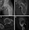
Radiographic appearance of fibrous dysplasia (FD). A) A proximal femur with typical ground glass appearance and shepherd's crook deformity in a 10-year-old child is shown. B) The appearance of FD in the femur of an untreated 40-year-old man demonstrates the tendency for FD to appear more sclerotic with time C) The typical ground glass appearance of FD in the craniofacial region on a CT image of a 10-year-old child is shown. The white arrows indicate the optic nerves, which are typically encased with FD. D) A CT image in a 40-year-old woman demonstrates the typical appearance of craniofacial FD in an older person, with mixed solid and "cystic" lesions. The Hounsfield Unit measurements of "cystic" lesions are quite useful in distinguishing soft tissue "cystic" lesions from true fluid-filled cysts, which are much more uncommon and tend to behave aggressively with rapid expansion and compression of vital structures. E-G) Bone Scintigraphy in FD. Representative 99Tc-MDP bone scans which show tracer uptake at affected skeletal sites, and the associated skeletal disease burden score (see ref. Collins, 2005) are shown. E) A 50-year-old woman with monostotic FD confined to a single focus involving contiguous bones in the craniofacial region. F) A 42-year-old man with polyostotic FD shows the tendency for FD to be predominantly (but not exclusively) unilateral, and to involve the skull base and proximal femur. G) A 16-year-old boy with McCune-Albright syndrome and involvement of virtually all skeletal sites (panostotic) is shown [65].
Figure 3
Figure 3
Representative histological images of FD. A) Calvarial FD lesions are characterized by uninterrupted networks of bone trabeculae (b) embedded in the fibrous tissue (ft). B) In FD lesions from gnathic bones, newly formed bone trabeculae (b) are deposited within the fibrous tissue (ft) in a typical discontinuous and parallel pattern. C) Collagen fibers perpendicularly oriented to forming bone surfaces (Sharpey fibers, arrows) represent a recurrent histological feature of FD at all skeletal sites. D-E) Osteomalacic changes and FGF23 production in FD. D) In many cases of FD, processing for undecalcified embedding reveals excess osteoid (asterisks) and severe undermineralization of the fibrous dysplastic bone. E) The mineralization defect of the FD tissue is related to elevated levels of FGF23 produced by activated FD osteogenic cells (arrows), as shown by in situ hybridization.
Figure 4
Figure 4
Fracture rates in FD. Fractures rates (reported as mean number of fractures per patient per year) are shown. Fractures are more frequent in childhood, with the highest rate occurring between 6–10 years of age. While fractures do lessen after childhood, there is a persistent rate into adulthood [18].
Figure 5
Figure 5
Molecular defect and phenotype in McCune-Albright syndrome (MAS). The hormones MSH (melanocyte stimulating hormone), LH (luteinizing hormone), TSH (thyroid stimulating hormone), GHRH (growth hormone stimulating hormone), and ACTH (adrenocrotical stimulating hormone) all signal through the G protein (alpha, beta, gamma subunits) pathway. In MAS, the alpha subunit is mutated in such a way as to induce constitutive activation of adenylate cyclase, and thus produce high levels of intracellular cAMP. This results in increased production of melanin, estradiol (E2), testosterone (T), thyroxine (T4), growth hormone (GH), and cortisol. Dysregulated production of these hormones results in café-au-lait spots, precocious puberty, fibrous dysplasia, acromegaly, hyperthyroidism, and Cushing's disease, depending on the tissue harboring the somatic mutation.
Figure 6
Figure 6
Molecular and developmental defect in McCune-Albright syndrome (MAS). A sporadic mutation occurs in a single cell (bright spot) at some point early in development. If this occurs at the inner call mass stage (embryonic stem cell stage), tissues from all 3 germ layers will be affected. As the cells derived from this mutated clone are dispersed throughout the organism, the final phenotype emerges, MAS.
Figure 7
Figure 7
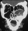
Fluid-filled cyst in FD. True fluid-filled cysts can occur in FD lesions. Shown is the CT scan of fluid-filled cyst that arose in a 12 year old boy with MAS who presented with facial nerve parasthesias and displacement of the orbit that occurred over approximately one week. The fluid-filled nature of the lesion is demonstrated by the fluid-fluid level (arrow). These lesions are more frequent in the craniofacial bones and can be aggressive. They usually require prompt surgical intervention.
Figure 8
Figure 8
CT for detecting subtle fractures. A) This radiograph was from a 9-year-old boy who complained of new-onset, focal pain in the groin. No discernable fracture is apparent. B) Reformatted CT images of the lesion revealed a fracture in the medial proximal femur (arrows).
Figure 9
Figure 9
Appearance of Cushing in a neonate. Rounded (moon) facies with plethora and facial hirsutism are present. This is usually accompanied by the presence of typical café-au-lait spots elsewhere on the examination.

References

    1. McCune DJ. Osteitis fibrosa cystica: the case of a nine-year-old girl who also exhibits precocious puberty, multiple pigmentation of the skin and hyperthyroidism. Am J Dis Child. 1936;52:743–744.
    1. Albright F, Butler AM, Hampton AO, Smith P. Syndrome characterized by osteitis fibrosa disseminata, areas, of pigmentation, and endocrine dysfunction, with precocious puberty in females: report of 5 cases. N Engl J Med. 1937;216:727–746.
    1. Mastorakos G, Mitsiades NS, Doufas AG, Koutras DA. Hyperthyroidism in McCune-Albright syndrome with a review of thyroid abnormalities sixty years after the first report. Thyroid. 1997;7:433–439. - PubMed
    1. Sherman SI, Ladenson PW. Octreotide therapy of growth hormone excess in the McCune-Albright syndrome. Journal of endocrinological investigation. 1992;15:185–190. - PubMed
    1. Akintoye SO, Chebli C, Booher S, Feuillan P, Kushner H, Leroith D, Cherman N, Bianco P, Wientroub S, Robey PG, Collins MT. Characterization of gsp-mediated growth hormone excess in the context of McCune-Albright syndrome. J Clin Endocrinol Metab. 2002;87:5104–5112. doi: 10.1210/jc.2001-012022. - DOI - PubMed

MeSH terms