. 2008 Jun;36(11):3765-80.
doi: 10.1093/nar/gkn120. Epub 2008 May 19.

Structural polymorphism within a regulatory element of the human KRAS promoter: formation of G4-DNA recognized by nuclear proteins

Affiliations

Structural polymorphism within a regulatory element of the human KRAS promoter: formation of G4-DNA recognized by nuclear proteins

Susanna Cogoi et al. Nucleic Acids Res. 2008 Jun.

Abstract

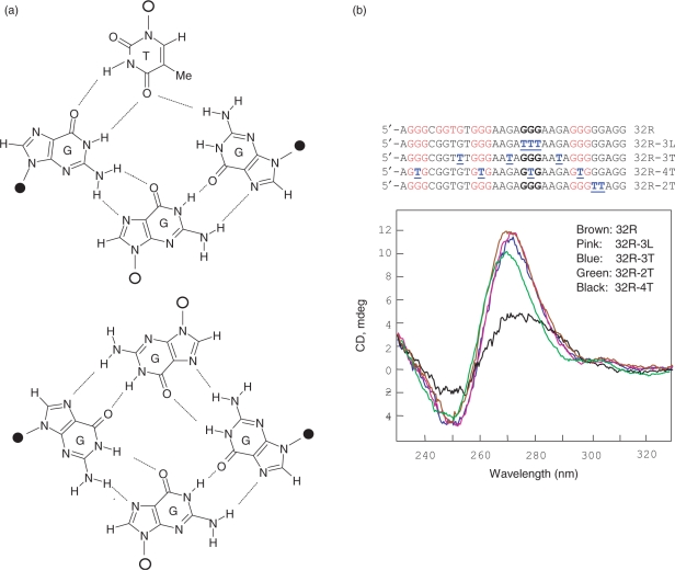
The human KRAS proto-oncogene contains a critical nuclease hypersensitive element (NHE) upstream of the major transcription initiation site. In this article, we demonstrate by primer-extension experiments, PAGE, chemical footprinting, CD, UV and FRET experiments that the G-rich strand of NHE (32R) folds into intra-molecular G-quadruplex structures. Fluorescence data show that 32R in 100 mM KCl melts with a biphasic profile, showing the formation of two distinct G-quadruplexes with T(m) of approximately 55 degrees C (Q(1)) and approximately 72 degrees C (Q(2)). DMS-footprinting and CD suggest that Q(1) can be a parallel and Q(2) a mixed parallel/antiparallel G-quadruplex. When dsNHE (32R hybridized to its complementary) is incubated with a nuclear extract from Panc-1 cells, three DNA-protein complexes are observed by EMSA. The complex of slower mobility is competed by quadruplex 32R, but not by mutant oligonucleotides, which cannot form a quadruplex structure. Using paramagnetic beads coupled with 32R, we pulled down from the Panc-1 extract proteins with affinity for quadruplex 32R. One of these is the heterogeneous nuclear ribonucleoprotein A1, which was previously reported to unfold quadruplex DNA. Our study suggests a role of quadruplex DNA in KRAS transcription and provides the basis for the rationale design of molecular strategies to inhibit the expression of KRAS.


Figures

Figure 1.
Figure 1.
Polymerase stop assay. Lanes 1–4 show sequencing reactions by the dideoxy method. Primer extension reactions were performed in the Klenow buffer, in the presence of 100 mM NaCl (lane 5), 100 mM KCl (lanes 6 and 8), 100 mM KCl plus 100 nM TMPyP4 (T) (lanes 7 and 9). Two DNA polymerase pauses are observed in the presence of KCl plus TMPyP4. The exact positions at which DNA polymerase I is arrested are indicated with arrows. As DNA template for the primer extension reactions we used a plasmid containing KRAS NHE. The primer extention reactions have been performed after incubation of the samples for 1 or 3 h at 50°C.
Figure 2.
Figure 2.
(a) Thermal differential spectra of 3 μM 32R (black open triangle) and CMYC (red open diamond) (CMYC sequence in Table 1) in 50 mM Tris–HCl, pH 7.4, 100 mM KCl. The spectrum of 3 μM 32R (blue filled cirlce) in 50 mM Tris–HCl, pH 7.4, 100 mM LiCl is also shown; (b) circular dichroism spectra as a function of temperature of 3 μM 32R in 50 mM Tris–HCl, pH 7.4, 100 mM KCl; spectra from top to bottom have been obtained at 20, 30, 40 50, 60, 65, 70, 75, 80, 90; (c) 15% non-denaturing PAGE (TBE 1×, 50 mM KCl in the gel and running buffer) of 20 nM 32P-labelled 32R, dsNHE (32R hybridized to complementary 32Y), 32Y and 32deg (32ss indicates a single-stranded unstructured 32-mer oligonucleotide), incubated overnight in 50 mM Tris–HCl, pH 7.4, 10 mM MgCl2, 140 mM KCl (buffer A) as follows. Lane 1: 32R in buffer A, heated at 95°C just before loading; lane 2: 32R added with cold 32R up to 1 μM in buffer A, heated at 95°C and incubated at 37°C for 24 h before loading; lanes 3 and 4: 32R+5 eq. TMPyP4 (T) incubated overnight (at 37 or 4°C) in buffer A; lanes 5 and 6: 32R+5 eq. TMPyP4 (T) incubated overnight (37°C or 4°C) in 50 mM Tris–HCl, pH 7.4, 10 mM MgCl2, 50 mM KCl; lanes 7–9: dsNHE, 32Y and 32deg incubated overnight in buffer A; (d) 15% non-denaturing PAGE performed as in (c) but with Li+ instead of K+.
Figure 3.
Figure 3.
Fluorescence emission spectra (500–650 nm) of 300 nm dual-labelled 32R incubated at 37°C in 50 mM Tris–HCl, pH 7.4, at different LiCl (a) or KCl (b) concentrations (10, 20, 40, 60 and 100 mM, the arrow indicates the KCl(or LiCl)-increment direction). Excitation wavelength was 475 nm; (c) First derivative fluorescence melting curves (dF/dT versus T) of 300 nm dual-labelled 32R in 50 mM Tris–HCl, pH 7.4, at different KCl concentrations (10, 20, 40, 60, 100 mM). Before measurements, the samples were incubated overnight in the appropriate buffer.
Figure 4.
Figure 4.
(a) dF/dT versus T melting curves of 300 nM dual-labelled 32R in 50 mM Tris–HCl, pH 7.4, 100 mM KCl at TMPyP4/32R ratios (r) of 0, 1, 3 and 5; (b) CD spectra of 32R in 50 mM Tris–HCl, pH 7.4, 100 mM KCl, room temperature, in the presence and absence of 5 eq. of TMPyP4. The oligonucleotide concentration was 3 μM, pathlength cell 0.5 cm, ellipticity expressed in millidegrees.
Figure 5.
Figure 5.
(a, left) DMS-footprinting of 32R at 37°C in 50 mM sodium cacodylate, pH 8, 1 mM EDTA 1 μg sonicated salmon sperm DNA (buffer B). 32R incubated overnight in water (lane 1); 32R incubated overnight in buffer B plus 100 mM LiCl (lane 2); 32R incubated in buffer B plus 50 or 140 mM KCl, 37°C, (lanes 3 and 4); 32R incubated in buffer B plus 50 or 140 mM KCl + 5 eq. TMPyP4 (lanes 5 and 6); 32R in buffer B plus 50 or 140 mM KCl, 50°C, (lanes 7 and 8); (a, right) 32R in buffer B plus 140 nM KCl (lane 10); 32R incubated overnight in buffer B, 100 mM KCl and 5 eq. of TMPyP4, irradiated for 5, 10 and 15 min (lanes 11–13);.(b) The proposed G-quadruplex structures for 32R inferred from footprinting studies. The circles in Q1 and Q2 indicate the bases of the loops: red, guanine; blue, A or T or C. Guanine 25 in Q2 is shown in red to indicate that is strongly photocleaved by TMPyP4. The expected quadruplex Qo is not supported by DMS-footprinting. The symbols above the sequence at the top mean: cleaved base (full square); uncleaved base (open square); partially cleaved base (semi-full square).
Figure 6.
Figure 6.
(a) Structures of the putative GGGT and GGGG tetrads; (b) CD spectra of 32R mutant sequences in 50 mM Tris–HCl, pH 7.4, 100 mM KCl. Oligonucleotide concentration was 3 μM, pathlength cell 0.5 cm, ellipticity expressed in millidegrees.
Figure 7.
Figure 7.
Competition binding experiments. When 32P-labelled dsNHE is incubated for 2 h at 37°C with a nuclear protein extract from Panc-1 cells, two main DNA–protein complexes (C1 and C3) are observed in a 5% PAGE in TBE at room temperature (a) The formation of the DNA–protein complex C1 is competed by 32R, but not by its mutant sequences 32R-4C, 32R-8C and 32R-12C (their sequences are in Table 1), that are unable to assume a G-quadruplex conformation, added to the reaction mixtures in excess over duplex dsNHE. Lanes 2–4: radiolabelled dsNHE (5 nM), 2 μg extract, 32R (competitor) 10-, 50-, 100-fold in excess respect dsNHE, respectively; lanes 5–7: same loading but with 32R-4C as competitor; lanes 8–10: same loading but with 32R-8C as competitor; lanes 11–13: same loading but with 32R-12C as competitor (b) When 32P-labelled 32R is incubated for 2 h at 37°C with the Panc-1 nuclear extract, three major DNA–protein complexes, B1, B2 and B3 are observed by 5% PAGE. The DNA–protein complex B1 is partially competed by the mutant oligonucleotides 32R-4T (30×, lane 2), 32R-8C (20× and 30×, lanes 3 and4), 32R-12C (30× lane 5), dsNHE (30×, lane 7). Complexes B2 and B3 are not competed by the mutant oligonucleotides, but they are competed by dsNHE.
Figure 8.
Figure 8.
(a) SDS–PAGE/silver staining of protein fractions eluted from streptavidin–biotin paramagnetic beads coupled to dsNHE (lanes 5–7) or to quadruplex 32R (lanes 2–4). Bands in the boxes have been excised and subjected to mass spectrometry analysis; (b, left) Southwestern blot showing that radiolabelled 32R-quadruplex, but not mutant 32R-4C, recognizes 34 and 70 kDa proteins (hnRNP A1 and Ku70) present in the extract; (b, right) Southwestern blot showing that radiolabelled 32R recognizes bands at 114 and 70 kDa (PARP-1 and Ku70) present in the 0.75 M NaCl pull-down fraction (lane 4); as a control it is shown that 32R recognizes recombinant PARP-1 (rPARP-1).
Figure 9.
Figure 9.
Real-time PCR determination of the level of endogenous KRAS mRNA in Panc-1 cells untreated or treated for 12 and 24 h with 50 μM of TMPyP2, TMPyP3 and TMPyP4. The ordinate reports the percent residual KRAS mRNA [T/C*100, where T is (RAS mRNA/GAPDH mRNA) in treated cells and C is (RAS mRNA/GAPDH mRNA) in untreated cells]. The structures of the three positional isomers TMPyP2, TMPyP3 and TMPyP4, in which the N-methyl group on the pyridyl ring is either in the ortho or meta or para positions, relative to its connection to the porphine core, are shown.

Similar articles

Cited by

References

    1. Barbacid M. Ras genes. Annu. Rev. Biochem. 1987;56:779–827. - PubMed
    1. Malumbres M, Barbacid M. RAS oncogenes: the first 30 years. Nat. Rev. Cancer. 2003;3:459–465. - PubMed
    1. Yanes L, Groffen J, Valenzuela DM. cKRAS mutations in human carcinomas occur preferentially in codon 12. Oncogene. 1987;1:315–318. - PubMed
    1. Nagata Y, Abe M, Kobayashi K, Yoshida K, Ishibashi T, Naoe T, Nakayama E, Shiku H. Glycine to aspartic acid mutations at codon 13 of the c-Ki-ras gene in human gastrointestinal cancers. Cancer Res. 1990;50:480–482. - PubMed
    1. Adjei AA. Blocking oncogenic Ras signaling for cancer therapy. J. Natl Cancer. Inst. 2002;94:1031–1032. - PubMed

Publication types