. 2008 Jul;28(14):4469-79.
doi: 10.1128/MCB.01416-07. Epub 2008 May 19.

Phosphorylation of threonine 61 by cyclin a/Cdk1 triggers degradation of stem-loop binding protein at the end of S phase

Affiliations

Phosphorylation of threonine 61 by cyclin a/Cdk1 triggers degradation of stem-loop binding protein at the end of S phase

M Murat Koseoglu et al. Mol Cell Biol. 2008 Jul.

Abstract

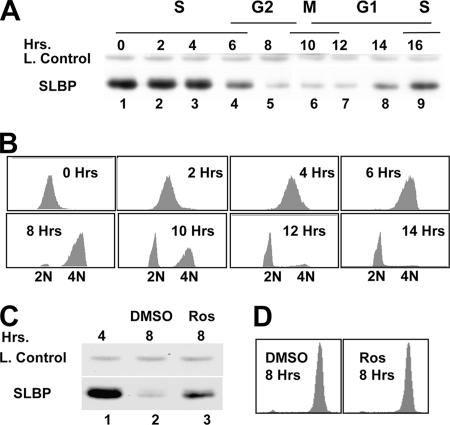
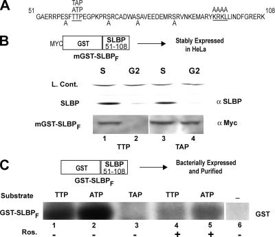
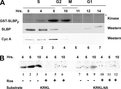
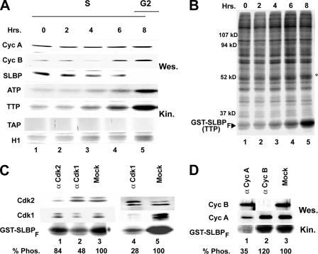
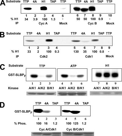
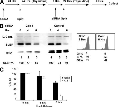
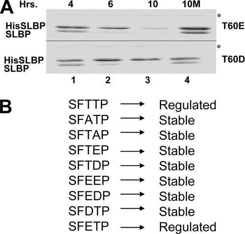
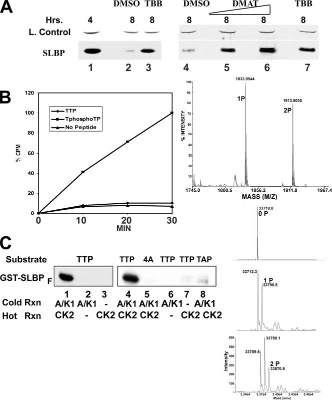
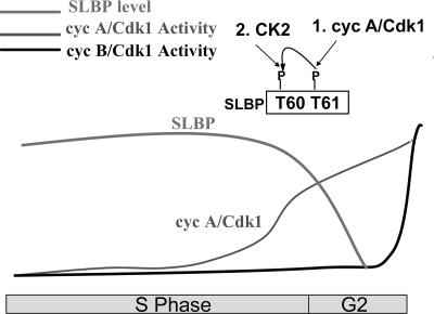
Histone mRNA levels are cell cycle regulated, and a major regulatory mechanism is restriction of stem-loop binding protein (SLBP) to S phase. Degradation of SLBP at the end of S phase results in cessation of histone mRNA biosynthesis, preventing accumulation of histone mRNA until SLBP is synthesized just before entry into the next S phase. Degradation of SLBP requires an SFTTP (58 to 62) and KRKL (95 to 98) sequence, which is a putative cyclin binding site. A fusion protein with the 58-amino-acid sequence of SLBP (amino acids 51 to 108) fused to glutathione S-transferase (GST) is sufficient to mimic SLBP degradation at late S phase. Using GST-SLBP fusion proteins as a substrate, we show that cyclin A/Cdk1 phosphorylates Thr61. Furthermore, knockdown of Cdk1 by RNA interference stabilizes SLBP at the end of S phase. Phosphorylation of Thr61 is necessary for subsequent phosphorylation of Thr60 by CK2 in vitro. Inhibitors of CK2 also prevent degradation of SLBP at the end of S phase. Thus, phosphorylation of Thr61 by cyclin A/Cdk1 primes phosphorylation of Thr60 by CK2 and is responsible for initiating SLBP degradation. We conclude that the increase in cyclin A/Cdk1 activity at the end of S phase triggers degradation of SLBP at S/G(2).


Figures

FIG. 1.
FIG. 1.
S/G2 degradation of SLBP is roscovitine sensitive. (A) Western blot analysis of SLBP levels in HeLa cells synchronized by a double-thymidime block and collected at the indicated time points after the release. (B) FACS analysis of the cells collected at indicated times after the release. (C) Western blot analysis of SLBP levels in HeLa cells which were treated with either dimethyl sulfoxide (DMSO, lane 2) or 20 μM roscovitine (Ros, lane 3) in DMSO 4 h after release from the double-thymidine block and collected four more hours later. (D) FACS analysis of both Ros- and DMSO-treated cells at the time of collection.
FIG. 2.
FIG. 2.
A fragment of SLBP fused to GST is sufficient to mimic S/G2 regulation of SLBP. (A) The SLBP fragment (amino acids 51 to 108) sufficient for S/G2 regulation is shown with the regions required for late-S-phase degradation underlined. The Ser/Thr residues that have been changed to alanine are shown by A below the wild-type sequence. Mutations that have been introduced in different constructs are indicated above the wild-type sequence. (B) HeLa cells stably expressing the Myc-GST-SLBP fragment (mGST-SLBPF) with wild-type TTP or TAP were synchronized by a double-thymidine block and collected at S or G2 phase. The levels of mGST-SLBPF and endogenous SLBP were determined by Western blot analysis using anti-Myc and anti-SLBP (α-Myc and α-SLBP) antibodies. (C) In vitro kinase assays with bacterially expressed GST-SLBPF (TTP, ATP, and TAP) or GST alone were performed in the presence of [γ-32P]ATP, using lysate from late-S-phase cells. The effect of roscovitine (Cdk1/Cdk2 inhibitor; 10 μM) on phosphorylation was also determined. Samples were analyzed by SDS-PAGE, and the phosphorylation level of each substrate was determined by autoradiography.
FIG. 3.
FIG. 3.
Lysates from late-S-phase cells phosphorylate GST-SLBPF in a KRKL-dependent and roscovitine-sensitive manner. (A) In vitro kinase assays were performed in the presence of [γ-32P]ATP, using lysates from HeLa cells collected at indicated time points after the double-thymidine block (top row). Equal amounts of protein from total cell lysates were used in each lane. Samples were analyzed by SDS-PAGE, and the phosphorylation level of the substrate was determined by autoradiography (top row). The levels of SLBP and cyclin A were determined by Western blot analysis using anti-SLBP and anti-cyclin A (CycA) antibodies (middle and bottom rows). The asterisk indicates a closely migrating protein in the lysate whose phosphorylation level does not show significant change throughout the cell cycle. (B) The effects of roscovitine (10 μM) and the KRKL region on phosphorylation of mGST-SLBPF were analyzed. The GST-SLBPF protein with either the wild-type KRKL region or the 4A mutation was used as a substrate (indicated below the panel).
FIG. 4.
FIG. 4.
Phosphorylation in lysates from late-S-phase cells is on Thr61. (A) In vitro kinase assays with the indicated form of mGST-SLBPF (ATP, TTP, or TAP) or histone H1 protein were performed in the presence of [γ-32P]ATP, using equal amounts of lysate from HeLa cells collected at indicated time points after a double thymidine block (rows 4, 5, 6, and 7). Samples were analyzed by SDS-PAGE, and the phosphorylation level of each substrate was determined by autoradiography (rows 4, 5, 6, and 7). Levels of cyclin A (Cyc A), cyclin B (Cyc B), and the SLBP protein in the lysates were examined by Western blot analysis using corresponding antibodies (rows 1, 2, and 3). (B) An autoradiograph of the entire gel is shown for the experiment in panel A, row 5. The asterisk indicates an endogenous protein that is phosphorylated in parallel with GST-SLBPF. The positions of molecular mass markers are shown. (C) Late-S-phase HeLa cell lysates were immunodepleted with anti-Cdk1 (lanes 2), anti-Cdk2 (lane 1), or protein A beads (lanes 3 and 5) or double immunodepleted with anti-Cdk1 (lane 5). The levels of Cdk2 (top) and Cdk1 (middle) in each lysate were determined by Western blot analysis with corresponding antibodies. The autoradiogram of the in vitro kinase assay using the depleted extracts and GST-SLBPF as a substrate is shown at the bottom. (D) In vitro kinase assay using late-S-phase HeLa cell lysates immunodepleted with anti-cyclin A (lane 1), anti-cyclin B (lane 2), or protein A beads (lane 3). The level of cyclin B (top) or cyclin A (middle) remaining in each lysate was determined by Western blot (Wes.) analysis with corresponding antibodies. The autoradiogram of the in vitro kinase assay (Kin.) using the depleted extracts and GST-SLBPF as a substrate is shown at the bottom. The phosphorylation level (Phos.) of each substrate in panels C and D was quantified using a PhosphorImager and indicated as a percentage below the autoradiograph.
FIG. 5.
FIG. 5.
The KRKL region is required for cyclin A/Cdk1 to phosphorylate Thr61. (A) Cyclin A (Cyc A) (lanes 1 to 4) and cyclin B (Cyc B) (lanes 6 to 9) immunoprecipitates (IP) from late-S-phase HeLa cell lysate were used in in vitro kinase assays in the presence of [γ-32P]ATP. Phosphorylation on the GST-SLBPF proteins TTP (lanes 1, 5, 6, and 10), KRKL/4A (lanes 2 and 7), TAP (lanes 4 and 9), and histone H1 (lanes 3 and 8) was detected by autoradiography. The phosphorylation level of each substrate was quantified using a PhosphorImager. For each set of reactions, the activity relative to that of histone H1 (set at 100) is given. (B) Cdk2 (lanes 1 to 4) or Cdk1 (lanes 5 to 8) immunoprecipitates from late-S-phase HeLa cell lysate were used in in vitro kinase assays in the presence of [γ-32P]ATP. Phosphorylation of GST-SLBPF proteins TTP (lanes 1, 5, and 9), KRKL/4A (lanes 2 and 6), and TAP (lanes 4 and 8), and of histone H1 (lanes 3 and 7) was detected by autoradiography. The phosphorylation level of each substrate was quantified by a PhosphorImager and normalized against the level of histone H1 phosphorylation. In the mock lanes (panel A, lanes 5 and 10, and panel B, lane 9), immunoprecipitations were done with just protein A beads. (C) Recombinant cyclin A/Cdk1 (lanes 1, 4, and 7), cyclin A/Cdk2 (lanes 2, 5, and 8), or cyclin B/Cdk1 (lanes 3, 6, and 9) were incubated with GST-SLBPF TTP (lanes 1 to 3) or ATP (lanes 4 to 6) or histone H1 (lanes 7 to 9) and [γ-32P]ATP. The proteins were resolved by gel electrophoresis and the phosphorylated proteins detected by autoradiography. The stained gel is shown below the lane. The experiment shown corresponds to a single gel and corresponding film image, with the assays with the different substrates separated by several lanes. (D) Recombinant cyclin A/Cdk1 (lanes 1 to 3) or cyclin B/Cdk1 (lanes 4 to 6) was incubated with GST-SLBPF TTP (lanes 1 and 4), KRKL/4A (lanes 2 and 5), or TAP (lanes 3 and 6). The proteins were resolved by gel electrophoresis and the phosphorylated (Phos.) proteins detected by autoradiography and quantified with a PhosphorImager. The activity was set at 100 for TTP with a particular kinase. The stained gel is shown below the lane. The experiment shown corresponds to a single gel and corresponding film image, with the assays with the different kinases separated by several lanes.
FIG. 6.
FIG. 6.
Cdk1 knockdown inhibits late-S-phase SLBP degradation. (A) Outline of experimental setup for combining RNAi treatment with the synchronization procedure. (B) HeLa cells were transfected with Cdk1 siRNA (lanes 1 to 3) or a control siRNA (Cont.) (lanes 4 to 6), C2, followed by synchronization with a double thymidine block. Cells were collected at the indicated time points after the release from the double thymidine block. The levels of Cdk1 and SLBP were determined by Western blot analysis with corresponding antibodies. A cross-reacting band detected by the SLBP antiserum serves as a loading control. FACS analysis of the 6-h time point from cells in panel B with percentage values for each cell cycle phase indicated is shown at the right. (C) The effects of the RNAi treatment on SLBP levels, determined by densitometry of the Western blots from two independent experiments, were averaged.
FIG. 7.
FIG. 7.
Changing Thr60 to Glu mimics phosphorylation of Thr60. (A) The first threonine, T60, in the SFTTP motif was changed to either glutamic acid (top) or aspartic acid (bottom) and stably expressed in HeLa cells as His-tagged SLBP (HisSLBP). Cells were synchronized by a double thymidine block and collected at the indicated hours after release. Levels of exogenous mutated His-tagged SLBP and endogenous SLBP were determined by Western blot analysis using SLBP antibody. The cells in lane 4 (10 M) were treated with MG132 (proteosome inhibitor) at 4 h after release and harvested at 10 h. (B) Summary of the effect of different mutations in the TTP motif on G2 SLBP degradation. “Regulated” indicates they showed a G2 degradation profile similar to that of endogenous SLBP.
FIG. 8.
FIG. 8.
CK2 phosphorylates Thr60 of SLBP. (A) Western blot analysis of SLBP levels in HeLa cells which were treated 4 h after release from a double thymidine block (at late S phase) and collected 4 h later at G2 with either DMSO (lanes 2 and 4) or different concentrations of two different CK2 inhibitors, TBB (75 μM [lane 3] or 50 μM [lane 7]) or DMAT (20 μM [lane 5] or 30 μM [lane 6]). The loading control is a cross-reacting band detected by the SLBP antisera. Lane 1 is a Western blot of cells 4 h after release prior to treatment. (B) The synthetic peptide TTP (54RRPESFTTPEGPKPR68) or the same peptide with a phosphothreonine at Thr 61 (TphosTP) was incubated with recombinant CK2 and [γ-32P]ATP, the peptides were bound to DEAE-paper, and the amount of the phosphorylated peptide was determined by liquid scintillation counting. The results are the averages from two experiments. On the right is analysis of the peptide by MALDI-TOF after 30 min of incubation with CK2. (C) The indicated GST-SLBPF substrates (above each lane) were incubated with unlabeled ATP and cyclin A/Cdk1 (lanes 1, 2, 4, 6, and 8) or buffer (lanes 3 and 7) overnight at 30°C. After the first incubation, CK2 (lanes 1, 3 to 5, 7, and 8) or buffer (lanes 2 and 6) was added together with [γ-32P]ATP and incubation continued for 30 min. The proteins were resolved by gel electrophoresis and the phosphorylated GST-SLBPF protein detected by autoradiography. GST-SLBPF was also incubated with unlabeled ATP and cyclin A/Cdk1 followed by CK2. GST-SLBPF was analyzed by electrospray ionization mass spectrometry, and the deconvoluted spectra of GST-SLBPF are shown (right, top), as is GST-SLBPF after incubation with cyclin A/Cdk1 (right, middle) and after incubation with cyclin A/Cdk1 followed by incubation with CK2 (right, bottom). The new peaks in the middle and bottom panels correspond to addition of one and two phosphates, respectively. Rxn, reaction.
FIG. 9.
FIG. 9.
Model for triggering SLBP degradation at S/G2. In contrast to cyclin B/Cdk1 activity, which increases rapidly at G2/M, there is a gradual increase in cyclin A/Cdk1 activity at the end of S phase that reaches a sufficient level at the S/G2 border to phosphorylate Thr61 of SLBP, and this primes subsequent phosphorylation of Thr60 by CK2 to mark SLBP for degradation.

Similar articles

Cited by

References

    1. Adams, P. D., X. T. Li, W. R. Sellers, K. B. Baker, X. H. Leng, J. W. Harper, Y. Taya, and W. G. Kaelin, Jr. 1999. Retinoblastoma protein contains a C-terminal motif that targets it for phosphorylation by cyclin-cdk complexes. Mol. Cell. Biol. 191068-1080. - PMC - PubMed
    1. Adams, P. D., W. R. Sellers, S. K. Sharma, A. D. Wu, C. M. Nalin, and W. G. Kaelin, Jr. 1996. Identification of a cyclin-cdk2 recognition motif present in substrates and p21-like cyclin-dependent kinase inhibitors. Mol. Cell. Biol. 166623-6633. - PMC - PubMed
    1. Archambault, V., N. E. Buchler, G. M. Wilmes, M. D. Jacobson, and F. R. Cross. 2005. Two-faced cyclins with eyes on the targets. Cell Cycle 4125-130. - PubMed
    1. Baldin, V., C. Cans, M. Knibiehler, and B. Ducommun. 1997. Phosphorylation of human CDC25B phosphatase by CDK1-cyclin A triggers its proteasome-dependent degradation. J. Biol. Chem. 27232731-32734. - PubMed
    1. Brown, N. R., M. E. Noble, J. A. Endicott, and L. N. Johnson. 1999. The structural basis for specificity of substrate and recruitment peptides for cyclin-dependent kinases. Nat. Cell Biol. 1438-443. - PubMed

Publication types