. 2008 Jul 25;283(30):20830-40.
doi: 10.1074/jbc.M801314200. Epub 2008 May 19.

Opalin, a transmembrane sialylglycoprotein located in the central nervous system myelin paranodal loop membrane

Affiliations

Opalin, a transmembrane sialylglycoprotein located in the central nervous system myelin paranodal loop membrane

Fumio Yoshikawa et al. J Biol Chem. .

Abstract

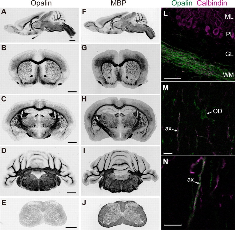
In contrast to compact myelin, the series of paranodal loops located in the outermost lateral region of myelin is non-compact; the intracellular space is filled by a continuous channel of cytoplasm, the extracellular surfaces between neighboring loops keep a definite distance, but the loop membranes have junctional specializations. Although the proteins that form compact myelin have been well studied, the protein components of paranodal loop membranes are not fully understood. This report describes the biochemical characterization and expression of Opalin as a novel membrane protein in paranodal loops. Mouse Opalin is composed of a short N-terminal extracellular domain (amino acid residues 1-30), a transmembrane domain (residues 31-53), and a long C-terminal intracellular domain (residues 54-143). Opalin is enriched in myelin of the central nervous system, but not that of the peripheral nervous system of mice. Enzymatic deglycosylation showed that myelin Opalin contained N- and O-glycans, and that the O-glycans, at least, had negatively charged sialic acids. We identified two N-glycan sites at Asn-6 and Asn-12 and an O-glycan site at Thr-14 in the extracellular domain. Site-directed mutations at the glycan sites impaired the cell surface localization of Opalin. In addition to the somata and processes of oligodendrocytes, Opalin immunoreactivity was observed in myelinated axons in a spiral fashion, and was concentrated in the paranodal loop region. Immunogold electron microscopy demonstrated that Opalin was localized at particular sites in the paranodal loop membrane. These results suggest a role for highly sialylglycosylated Opalin in an intermembranous function of the myelin paranodal loops in the central nervous system.


Figures

FIGURE 1.
FIGURE 1.
Structure of mouse Opalin protein and mRNA expression. A, schematic representation of mouse Opalin (1–143 aa). The predicted transmembrane domain (31–53 aa) is shown in the closed box. Two N-linked glycosylation sites at Asn-6 (N6) and Asn-12 (N12) are indicated by open triangles and an O-linked glycosylation site at Thr-14 (T14) is indicated by a closed triangle. Six predicted protein phosphorylation sites are indicated by vertical bars with single-letter amino acid positions: Thr-59 (T59) (cAMP-dependent protein kinase and protein kinase C), Tyr-78 (Y78) (insulin receptor), S90 (mitogen-activated protein kinase, cdc2, and cyclin-dependent kinase), Thr-92 (T92) (protein kinase A), Tyr-106 (Y106) (epidermal growth factor receptor and INSR), and Tyr-121 (Y121) (spleen tyrosine kinase and INSR). Three deletion mutants (dC, dIC, and dN) contained parts of the full-length structure (FL) (see supplemental Fig. S2). dC has a deletion of the C-terminal 38 aa (and thus comprises residues 1 to 115), dN has a deletion of the N-terminal 26 (and thus comprises residues 27 to 143), and dIC has a deletion of the intracellular domain from 55 to 115 aa (and thus comprises residues 1 to 54 and 116 to 143). B, RT-PCR analysis of developmental time series expression. Expression of Opalin, PLP/DM20 (two splice variants, upper band; PLP, lower band, DM20), MBP, MOG, and glyceraldehyde-3-phosphate dehydrogenase (GAPDH) mRNAs during the postnatal stages of mouse cerebellar development are shown. Opalin mRNA expression is up-regulated in the postnatal mouse cerebellum, similar to the expression of myelin-related genes. C, RT-PCR analysis of tissue specificity. Expression of Opalin mRNA in the brain, thymus, lung, heart, liver, spleen, kidney, and testis of mice at P21 is shown. Opalin mRNA expression is brain specific. D, in situ hybridization analysis of spatial cellular expression. Expression of Opalin mRNAs in sagittal brain sections of P21 mice is shown. Opalin mRNA is concentrated in white matter-rich regions, including the optic chiasm, corpus callosum, anterior commissure, and the white matter layer of the cerebellar cortex. ox, optic chiasm; cc, corpus callosum; df, dorsal fornix; ac, anterior commissure; ML, molecular layer; GL, granular layer; WM, white matter.
FIGURE 2.
FIGURE 2.
Western blot analysis of Opalin protein in subcellular fractions of the mouse nervous system. A, Western blot analysis of subcellular fractions from postnatal mouse cerebellum at P1, P7, P14, and P21 with the anti-Opalin antibody. Opalin, with a molecular mass of ∼33–36 kDa, is detected in precipitated membrane-rich fractions between P14 and P21. ppt1, precipitate nuclear fraction; ppt2 + 3, precipitate microsomal fraction; sup3, soluble cytosolic fraction. Molecular mass standards are indicated on the left. B, Western blot analysis of myelin fractions with the anti-Opalin antibody. Equal amounts of protein samples were loaded in each lane. Opalin was more enriched in the crude and pure myelin fractions than in ppt2 + 3, similar to the myelin marker MBP (bottom panel). C, Western blot analysis of CNS and PNS myelin fractions with the anti-Opalin antibody. Opalin protein is concentrated in CNS myelin fractions (from whole brain or spinal cords) and not in PNS myelin fractions (from sciatic nerves), whereas MBP is abundant in both fractions (bottom panel). The anti-MBP antibody reacts with multiple splice variants. Molecular mass standards are indicated on the left.
FIGURE 3.
FIGURE 3.
Immunohistochemical localization analysis of Opalin in mouse brains. A–J, sagittal (A and F) and coronal (BE and G–J) sections of P21 mouse brains were immunoreacted with anti-Opalin antibody (A–E) and anti-MBP antibody (F–J) and detected by HRP-DAB stain. B and G, regions including the frontal cortex and caudate-putamen; C and H, regions including the cerebral cortex, hippocampus and thalamus; D and I, regions including the cerebellum and brain stem; E and J, spinal cords. Opalin is predominantly localized in white matter-rich regions. L–N, sagittal sections of P14 mouse cerebellum were double-immunostained with anti-Opalin (green) and anti-calbindin (magenta) antibodies and were observed by fluorescent confocal microscopy. M and N, higher magnifications of the granular layer of the cerebellum. Opalin immunoreactivity is found in soma and process of oligodendrocytes (M) and is also localized in myelinating axons in a spiral fashion (N). ML, molecular layer; PL, Purkinje cell layer; GL, granular layer; WM, white matter; OD, oligodendrocyte cell body; ax, axon of Purkinje cells. Scale bars, 1 mm in A–D; 0.5 mm in E; 50 μm in L; 10 μm in M and N.
FIGURE 4.
FIGURE 4.
Enzymatic deglycosylation analysis of myelin Opalin and identification of N- and O-glycosylation sites at Asn-6, Asn-12, and Thr-14 residues of Opalin by site-directed amino acid substitutions. A, enzymatic deglycosylation of myelin fractions prepared from P23 mouse brains with N-glycosidase F (N-Glyc), O-glycosidase (O-Glyc), and neuraminidase (NA) followed by immunoblotting with anti-Opalin antibody. Faster mobility on SDS-PAGE upon deglycosylation treatment indicates the glycosylation of myelin Opalin; Opalin contains both N-linked and O-linked glycans, both types of which are sialylated. B, Western blot analysis of protein lysates from COS7 cells transfected with the Opalin wild-type (wt), Asn-6 to Gln substitution mutant (N6Q), Asn-12 to Gln substitution mutant (N12Q), Thr-12 to Ala substitution mutant (T14A), N6Q/N12Q double mutant, N6Q/N12Q/T14Q triple mutant, and vector alone. Myelin, myelin fraction of mouse brains. Opalin is N-glycosylated at Asn-6 and Asn-12 and O-glycosylated at Thr-14. The N6Q/N12Q/T14A mutant migrates along with N6Q/N12Q on SDS-PAGE, suggesting that the N6Q/N12Q mutation affects the O-glycosylation pattern. Enzymatic deglycosylation experiments indicated that the N6Q/N12Q/T14A mutant expressed in COS7 cells still contains glycans at other site(s) (supplemental Fig. S2). Molecular mass standards (in kDa) are shown on the left.
FIGURE 5.
FIGURE 5.
Opalin is localized to the cell surface and cell-cell contact sites in HeLa cells but the mutants lacking the glycan sites are mislocalized to the endoplasmic reticulum-Golgi network. C-terminal GFP fusion constructs of Opalin (A), N6Q (B), N12Q (C), T14A (D), N6Q/N12Q (E), N6Q/N12Q/T14A (F), and GFP alone (G) were transfected into HeLa fibroblasts. Panels A–G show GFP fluorescent images (green) in a 2-μm optical slice taken with a confocal microscope equipped with the ×63 objective lens. Panels H and I show GFP (green) and phalloidin-actin filament (magenta) fluorescent images of a 1-μm confocal optical slice around the bottom of the cells, observed using the ×100 objective lens. Wild-type Opalin was predominantly localized in spike-like protrusions (H and I) as well as the cell periphery and cell-cell contact sites (A) and was partly colocalized with phallodin-labeled actin filaments. On the other hand, the glycan mutants were concentrated in the endoplasmic reticulum-Golgi networks (stained with marker proteins, data not shown) rather than at the cell surface (B–F). In addition, these mutants, especially the N6Q/N12Q/T14A triple mutant, tended to induce large membrane protrusions (arrows in C–F). Scale bars, 10 μm in panels G and H and 5 μm in panel I.
FIGURE 6.
FIGURE 6.
Localization of transfected Opalin-Venus and N6Q/N12Q-Venus in primary cultured oligodendrocytes and immunolocalization of native Opalin in myelinating axons of spinal cords. A, Opalin-Venus and N6Q/N12Q-Venus were transfected into primary oligodendrocyte cultures prepared from rat cerebrum. Venus fluorescence was observed in O4-immunopositive cells extending multiple thick processes (data not shown), most of which were oligodendrocytes, as described previously (39). Opalin-Venus was localized in soma, processes, and thin membranous structures between processes. On the other hand, N6Q/N12Q-Venus was punctately distributed throughout the oligodendrocytes. B and C, the native Opalin in the myelinating axons of spinal cords from 8-week-old mice was immunolocalized together with neurofilament 160 (B, left panels), MAG (B, right), Na+ channel (C, left), and caspr (C, right). Opalin immunoreactivity localized to myelin-wrapped neurofilament-immunopositive axons in a spiral-like fashion was only partly colocalized with MAG immunoreactivity. Opalin immunoreactivity was more concentrated around the regions flanking the node. Scale bars, 5 μm.
FIGURE 7.
FIGURE 7.
Opalin is localized at specific sites in the paranodal and inner loop regions of mouse optic nerves. Immunogold electron microscopic analysis of the optic nerves of 2-week-old mice. A, Opalin immunogold particles in a transverse section through the optic nerve. Inset, magnification of the inner mesaxon area in the boxed area. Opalin immunogold particles are localized around the inner loop (or adaxonal) membrane (or inner mesaxon), and none are seen in the compact myelin or axo-glial junctions. The dotted line in the inset indicates the axo-glial junction. B and C, Opalin immunogold particles in longitudinal sections of optic nerves. Opalin immunogold particles are localized on the cytoplasmic side immediately below the paranodal loop membranes. Pairs of immunogold particles are often observed on apposing membranes. Arrows indicate the position of the immunogold particles. Ax, axon; CM, compact myelin; IL, inner loop; PN, paranode. Scale bars, 0.1 μm.

Similar articles

Cited by

References

    1. Roach, A., Boylan, K., Horvath, S., Prusiner, S. B., and Hood, L. E. (1983) Cell 34 799–806 - PubMed
    1. Matthieu, J. M., Roch, J. M., Omlin, F. X., Rambaldi, I., Almazan, G., and Braun, P. E. (1986) J. Cell Biol. 103 2673–2682 - PMC - PubMed
    1. Boison, D., and Stoffel, W. (1994) Proc. Natl. Acad. Sci. U. S. A. 91 11709–11713 - PMC - PubMed
    1. Rosenbluth, J., Nave, K. A., Mierzwa, A., and Schiff, R. (2006) Glia 54 172–182 - PubMed
    1. Morita, K., Sasaki, H., Fujimoto, K., Furuse, M., and Tsukita, Sh. (1999) J. Cell Biol. 145 579–588 - PMC - PubMed

Publication types