. 2008 Jun 9;205(6):1261-8.
doi: 10.1084/jem.20080108. Epub 2008 May 19.

"Re-educating" tumor-associated macrophages by targeting NF-kappaB

Affiliations

"Re-educating" tumor-associated macrophages by targeting NF-kappaB

Thorsten Hagemann et al. J Exp Med. .

Abstract

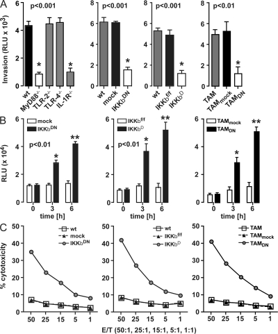
The nuclear factor kappaB (NF-kappaB) signaling pathway is important in cancer-related inflammation and malignant progression. Here, we describe a new role for NF-kappaB in cancer in maintaining the immunosuppressive phenotype of tumor-associated macrophages (TAMs). We show that macrophages are polarized via interleukin (IL)-1R and MyD88 to an immunosuppressive "alternative" phenotype that requires IkappaB kinase beta-mediated NF-kappaB activation. When NF-kappaB signaling is inhibited specifically in TAMs, they become cytotoxic to tumor cells and switch to a "classically" activated phenotype; IL-12(high), major histocompatibility complex II(high), but IL-10(low) and arginase-1(low). Targeting NF-kappaB signaling in TAMs also promotes regression of advanced tumors in vivo by induction of macrophage tumoricidal activity and activation of antitumor activity through IL-12-dependent NK cell recruitment. We provide a rationale for manipulating the phenotype of the abundant macrophage population already located within the tumor microenvironment; the potential to "re-educate" the tumor-promoting macrophage population may prove an effective and novel therapeutic approach for cancer that complements existing therapies.


Figures

Figure 1.
Figure 1.
IKKβ inhibition increases macrophage tumoricidal activity. (A) WT, MyD88−/−, TLR-2−/−, and TLR-4−/− or mock, IKKβDN or IKKβf/f, and IKKβΔ BMDMs were co-cultured with ID8-Luc in a modified Boyden chamber without direct cell–cell contact for 72 h. In addition, CD11b+-selected TAMs from the ascites of ID8 tumor–bearing mice were mock (TAMmock) or IKKβDN infected (TAMDN). Invasion of ID8-Luc cells was assessed by luciferase activity in the lower part of the chamber. Macrophages deleted in MyD88, IL-1R, or IKKβ significantly reduce ID8-Luc invasion (P < 0.01; t test with Welch's correction). Data are represented as mean ± SEM of n = 6. Representative data are shown from at least three independent experiments. (B) ID8 cells were co-cultured with IKKβ-targeted BMDMs or TAMs and respective control cells. Fluorescence caspase 3/7 activity was assessed after 0, 3, and 6 h. Co-culture with IKKβDN and IKKβΔ BMDMs or TAMs expressing IKKβDN (TAMDN) significantly increases caspase 3/7 activity in ID8 cells (P < 0.01; t test with Welch's correction). Data are represented as mean ± SD of n = 6. Representative data are shown from at least three independent experiments. (C) [111In]oxine release assay after a 24-h co-culture of 111In-labeled ID8 cells with mock, IKKβDN, TAMDN or IKKβf/f, and IKKβΔ macrophages. 111In-release in cell-free culture supernatants was measured after 24 h with a scintillation counter. IKKβ-targeted macrophages promote increased cytotoxicity. Data are represented as mean of n = 3. Representative data are shown from at least three independent experiments.
Figure 2.
Figure 2.
IKKβ maintains the alternative phenotype of tumor-polarized macrophages. WT, mock, IKKβDN or IKKβf/f, and IKKβΔ BMDMs were co-cultured with ID8 cells for 24 h. Supernatant was collected after 24 h and analyzed for IL-10, IL-12p70, TNF-α, and NO production by ELISA. (A) Co-cultured WT macrophages express an immunosuppressive IL-10high IL-12p70low TNF-αhigh profile. However, supernatant from co-cultured IKKβ-deleted macrophages show a significant decrease in IL-10 and TNF-α (P < 0.01; t test with Welch's correction) but increase in IL-12p70 (P < 0.01; t test with Welch's correction). Data are represented as mean ± SD of n = 3. Representative data are shown from at least three independent experiments. (B) In parallel, total RNA was isolated for real-time PCR analysis of IL-12p40 and arginase-1 in co-cultured macrophages. Targeting IKKβ in macrophages switches their phenotype to an IL-12high arginase-1low NOS2high profile, consistent with an M1 phenotype. In_addition, IKKβ-targeted macrophages secrete significantly higher levels of NO, measured as nitrite by Griess assay (P < 0.01; Mann-Whitney test). Data are represented as mean ± SD of n = 3. Representative data are shown from at least four independent experiments. (C) Macrophages were co-cultured with ID8 cells, and protein extracts were prepared at the indicated time points for biochemical analysis. Expression of NF-κB p65, serine 536 phosphorylation of p65, IKKβ, Stat1, tyrosine 701 phosphorylation of Stat1 (Tyr701), and NOS2 was measured by immunoblot analysis of cell lysates using β-actin as a loading control. Representative data are shown from at least three independent experiments. (D) [111In]oxine release assay after co-culture of ID8 ovarian cancer cells with IKKβDN or IKKβΔ BMDMs. IKKβ-targeted macrophages promote increased cytotoxicity that was reversed by the addition of the selective NOS2 inhibitor 1400W. Data are represented as mean of n = 3. Representative data are shown from at least three independent experiments.
Figure 3.
Figure 3.
Adoptive transfer of IKKβ-targeted BMDMs inhibits tumor growth in vivo. (A) Representative bioluminescence imaging in vivo 14 d after adoptive transfer of BMDMs (n = 6; three independent experiments). ID8-Luc were injected i.p. into syngeneic mice and tumors were allowed to develop. After 35 d, WT, mock, and IKKβDN BMDMs were adoptively transferred and peritoneal cells were collected after an additional 7 and 14 d. Bioluminescence is presented as a pseudocolor scale: red, the highest photon flux; blue, the lowest photon flux. (B) Quantification of bioluminescence from primary tumors (n = 6 each) obtained on days 0, 7, and 14. Adoptively transferred IKKβDN BMDMs significantly reduced ID8 growth on day 14 (P < 0.05; t test with Welch's correction). (C) NO secretion into the peritoneal cavity. IKKβDN BMDMs increased NO release 4 h after adoptive transfer (P < 0.05; t test with Welch's correction). Data are represented as mean ± SD of n = 6. Representative data are shown from at least two independent experiments. (D) Cytokine profile in tumor ascites after 14 d. Ascites from mice treated with IKKβ-targeted BMDMs contained significantly lower amounts of IL-10 and TNF-α but higher amounts of IL-12 (P < 0.01; Mann-Whitney test). Total RNA was isolated from CD11b+-selected ascitic macrophages for real-time PCR analysis of IL-12p40 and arginase-1 expression. Data are represented as fold induction of mRNA expression compared with WT macrophages (n = 6). (E) MHC class II expression in the ascitic CD11b+ myeloid cell population measured by FACS (percentage of positive cells indicated). Representative histograms are shown from n = 6.
Figure 4.
Figure 4.
Adoptive transfer of IKKβ-targeted TAMs inhibits tumor growth in vivo. (A) Representative bioluminescence imaging in vivo 14 d after adoptive transfer of TAMs isolated from established ID8 tumors (n = 6; three independent experiments). ID8-Luc were injected i.p. into syngeneic mice and tumors were allowed to develop. After 35 d, mock- and IKKβDN-infected TAMs (TAMmock, TAMDN, respectively) were adoptively transferred and peritoneal cells were collected after an additional 7 and 14 d. Bioluminescence is presented as a pseudocolor scale: red, the highest photon flux; blue, the lowest photon flux. (B) Quantification of bioluminescence from primary tumors (n = 6 each) obtained on days 0, 7, and 14. Adoptive transfer of TAMDN significantly reduced ID8 growth on day 14 (P < 0.05; t test with Welch's correction). (C) NO secretion into the peritoneal cavity. Transfer of TAMDN significantly increased NO production 4 h after adoptive transfer (P < 0.05; t test with Welch's correction). Data are represented as mean ± SD of n = 6. Representative data are shown from at least two independent experiments. (D) Cytokine profile in tumor ascites after 14 d. Ascitic fluid after transfer of TAMDN contained significantly lower amounts of IL-10 and TNF-α but higher amounts of IL-12 (P < 0.01; Mann-Whitney test). Total RNA was isolated from CD11b+-selected TAMs for real-time PCR analysis of IL-12p40 and arginase-1 expression. Data are represented as fold induction of mRNA expression compared with TAMmock-treated mice (n = 6). (E) MHC class II expression in the ascitic CD11b+ TAMs measured by FACS (percentage of positive cells indicated). Representative histograms are shown from n = 6.
Figure 5.
Figure 5.
Increased IL-12–dependent NK cell recruitment. (A) Neutralizing IL-12p40 but not IL-23p19 rescued the antitumor effect of adoptively transferred IKKβDN BMDMs (P < 0.01; Mann-Whitney test). Data are represented as mean ± SEM of n = 6. Representative data are shown from two independent experiments. (B) Representative bioluminescence image of the IL-12p40–neutralizing experiment (n = 6). (C) Adoptive transfer of IKKβDN BMDMs increases NK cell recruitment in ID8 tumors (P < 0.01; Mann-Whitney test). Data are represented as mean ± SD of n = 6. Representative data are shown from two independent experiments. (D) Ex vivo NK cell cytotoxicity assay. Splenic DX5+-enriched NK cells were stimulated with ascites from tumor-bearing mice, which had either no treatment or were treated with adoptive transfer of mock or IKKβDN BMDMs. Ascites from mice treated by adoptive transfer of IKKβDN BMDMs led to a significant increase in NK cell–mediated tumor cell cytotoxicity (P < 0.01; t test with Welch's correction). The addition of a neutralizing antibody against IL-12p40 but not IL-23p19 or the respective control antibody rescued the increased tumoricidal effect. Data are represented as mean of n = 3. Representative data are shown from three independent experiments.

Comment in

References

    1. Condeelis, J., and J.W. Pollard. 2006. Macrophages: obligate partners for tumor cell migration, invasion, and metastasis. Cell. 124:263–266. - PubMed
    1. Sica, A., T. Schioppa, A. Mantovani, and P. Allavena. 2006. Tumour-associated macrophages are a distinct M2 polarised population promoting tumour progression: potential targets of anti-cancer therapy. Eur. J. Cancer. 42:717–727. - PubMed
    1. Gordon, S. 2003. Alternative activation of macrophages. Nat. Rev. Immunol. 3:23–35. - PubMed
    1. Balkwill, F., K.A. Charles, and A. Mantovani. 2005. Smoldering and polarized inflammation in the initiation and promotion of malignant disease. Cancer Cell. 7:211–217. - PubMed
    1. Mantovani, A., A. Sica, S. Sozzani, P. Allavena, A. Vecchi, and M. Locati. 2004. The chemokine system in diverse forms of macrophage activation and polarization. Trends Immunol. 25:677–686. - PubMed

Publication types