. 2008 Jun 9;205(6):1269-76.
doi: 10.1084/jem.20080124. Epub 2008 May 19.

An antiinflammatory role for IKKbeta through the inhibition of "classical" macrophage activation

Affiliations

An antiinflammatory role for IKKbeta through the inhibition of "classical" macrophage activation

Carol Ho Yan Fong et al. J Exp Med. .

Abstract

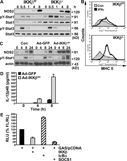
The nuclear factor kappaB (NF-kappaB) pathway plays a central role in inflammation and immunity. In response to proinflammatory cytokines and pathogen-associated molecular patterns, NF-kappaB activation is controlled by IkappaB kinase (IKK)beta. Using Cre/lox-mediated gene targeting of IKKbeta, we have uncovered a tissue-specific role for IKKbeta during infection with group B streptococcus. Although deletion of IKKbeta in airway epithelial cells had the predicted effect of inhibiting inflammation and reducing innate immunity, deletion of IKKbeta in the myeloid lineage unexpectedly conferred resistance to infection that was associated with increased expression of interleukin (IL)-12, inducible nitric oxide synthase (NOS2), and major histocompatibility complex (MHC) class II by macrophages. We also describe a previously unknown role for IKKbeta in the inhibition of signal transducer and activator of transcription (Stat)1 signaling in macrophages, which is critical for IL-12, NOS2, and MHC class II expression. These studies suggest that IKKbeta inhibits the "classically" activated or M1 macrophage phenotype during infection through negative cross talk with the Stat1 pathway. This may represent a mechanism to prevent the over-exuberant activation of macrophages during infection and contribute to the resolution of inflammation. This establishes a new role for IKKbeta in the regulation of macrophage activation with important implications in chronic inflammatory disease, infection, and cancer.


Figures

Figure 1.
Figure 1.
Tissue-specific role for IKKβ in Streptococcal pneumonia. (A) IKKβΔEpi and IKKβf/f-Otet-cre control mice were infected intranasally with 5 × 10⁶ CFUs GBS in PBS. Bronchialveolar lavage was performed after 4 h, and CFUs were determined by serial dilution on Todd-Hewitt agar plates (n = 11–14; *, P = 0.0242). (B) Hematoxylin and eosin stain (left, H+E) of lungs from GBS-infected mice after 4 h showed reduced leukocyte infiltration in IKKβΔEpi mice (arrows). Gram stain (right) of infected lungs showed accumulation of bacteria in alveolar spaces of IKKβΔEpi lungs (arrows). Representative panels are shown from n = 8. Bar, 100 μm. (C) IKKβΔMye and IKKβf/f control mice were infected as described above, and BAL CFUs were measured at 4 h (n = 11–14; *, P = 0.0304). (D) Hematoxylin and eosin stain of lungs from GBS-infected mice after 4 and 24 h showed resolution of peribronchial inflammation in the lungs of IKKβf/f mice at 24 h (top, arrows); however, inflammation in IKKβΔMye mice fails to resolve (bottom, arrows). Bar, 200 μm. (E) PMN recruitment in IKKβΔMye and IKKβf/f mice infected with GBS was counted under high power field after hematoxylin and eosin stain. Data are represented as mean ± SEM of n = 8 (**, P = 0.005).
Figure 2.
Figure 2.
IKKβ inhibits M1 macrophage activation during infection. (A) IKKβΔMye and IKKβf/f control mice were infected by intraperitoneal injection of 5 × 10⁷ CFUs GBS in PBS, and survival was monitored (n = 16–17). (B) Blood was collected at 4 h by retroorbital bleed. Serial dilutions of blood samples were plated in triplicate on Todd-Hewitt agar plates, and CFUs were counted (n = 9–10; **, P = 0.0057). (C) Serum levels of IL-12p70, IL-1β, and spleen IFN-γ were measured by ELISA (n = 7–8; IL-12p70: **, P = 0.0023; IFN-γ: *, P = 0.0148). (D) Total RNA was isolated from the spleens of GBS-infected mice for real-time PCR analysis of IL-12p40 and arginase-1 expression. Data are represented as fold induction of mRNA expression compared with uninfected mice (n = 7–8). (E) MHC class II expression on peritoneal macrophages measured by FACS (percentage of positive cells indicated). (F) Immunohistochemical analysis of NOS2 expression in GBS-infected lungs shows increased expression of NOS2 in alveolar macrophages of IKKβΔMye mice (arrows), whereas expression in bronchial epithelial cells remains unchanged (asterisks). Representative panels are shown from n = 8. Bar, 200 μm.
Figure 3.
Figure 3.
IKKβ inhibits M1 macrophage activation in response to LPS in vivo. (A) Peritoneal macrophages (PM) and (B) neutrophils (PMN) were isolated from IKKβΔMye and IKKβf/f mice for in vitro killing assays with GBS (MOI of 5:1). (C) IKKβΔMye and IKKβf/f mice were challenged intranasally with 10 ng LPS in PBS. After 4 h, lungs were harvested and digested for FACS analysis of CD11b (myeloid), Gr-1 (PMN), and MHC class II expression. A representative histogram of MHC class II expression on CD11b+ Gr-1 alveolar macrophages is shown. (D) Tabulated data of mean fluorescence intensity for CD11b+ Gr-1 MHC class II+–activated macrophages in the lung and PMN recruitment as percentage of BAL cells from LPS-challenged mice (data are represented as mean ± SEM of n = 6). (E) In parallel experiments, IL-12 and IL-10 levels were measured in lung homogenates from LPS-challenged mice by ELISA. Data are represented as mean ± SEM (n = 8; *, P = 0.0381). (F) Immunohistochemical analysis of NOS2 expression in LPS-challenged mice. IKKβΔMye mice show increased expression of NOS2 in alveolar macrophages compared with IKKβf/f control mice (arrows). Representative panels are shown from n = 6 mice. Bar, 100 μm.
Figure 4.
Figure 4.
IKKβ inhibits Stat-1 activation in GBS-infected macrophages. (A) Peritoneal macrophages from IKKβΔMye and IKKβf/f mice were stimulated in vitro with heat-killed GBS (MOI of 20:1). Protein extracts were prepared at the indicated time points for biochemical analysis. IKK and JNK activity was measured by IP kinase assay (KA) using recombinant substrates GST-IκBα1-54 or GST-cJun1-79, respectively. NF-κB activation was measured by electrophoretic mobility shift assay (EMSA) using 32P-labeled κB consensus oligonucleotide. (B) Expression of IKK, pro–IL-1β, NOS2, tyrosine 701 phosphorylation of Stat1 (pY-Stat1), serine 726 phosphorylation of Stat1 (pS-Stat1), total Stat1, and SOCS1 was measured by immunoblot analysis of cell lysates using actin as a loading control. (C) In parallel experiments, TNF-α production in cell culture supernatants was measured by ELISA, and (D) total RNA was isolated for real-time PCR analysis of TNF-α (Tnfa) and IFN-β (Ifnb) mRNA expression. Data are represented as mean ± SEM of n = 4. Representative data are shown from at least three independent experiments.
Figure 5.
Figure 5.
IKKβ inhibits IFN-γ signaling in macrophages. (A) Peritoneal macrophages from IKKβΔMye and IKKβf/f mice were stimulated in vitro with 100 ng/ml LPS in combination with 100 U/ml recombinant mouse IFN-γ, and protein extracts were prepared at the indicated time points. NOS2 expression, pY-Stat1, and pY-Stat3 were measured by immunoblot analysis. (B) Peritoneal macrophages from IKKβΔMye and IKKβf/f mice were stimulated in vitro, with IFN-γ and MHC class II expression analyzed by FACS. Representative data are shown from at least three independent experiments. (C) BMDMs were infected with recombinant adenovirus expressing a dominant-negative inhibitor of IKKβ (Ad-IKKβdn) or EGFP (Ad-GFP) 48 h before stimulation with LPS. NOS2 and pY-Stat1 expression was measured by immunoblot analysis at the indicated time points. (D) BMDMs were infected with Ad-IKKβdn or Ad-GFP before stimulation with LPS/IFN-γ, and IL-12p40 production was measured in cell culture supernatants by ELISA. Data are represented as mean ± SEM of n = 4. (E) RAW 264.7 macrophages were transfected with GAS luciferase reporter and either empty vector (pCDNA) or cDNA expressing IKKβ, IκBα, or SOCS1. After LPS/IFN-γ stimulation for 6 h, GAS reporter activity was measured by dual luciferase activity assay. Data are represented as relative light units (RLU) normalized for transfection efficiency with pRLTK.

Comment in

References

    1. Karin, M., and Y. Ben-Neriah. 2000. Phosphorylation meets ubiquitination: the control of NF-[kappa]B activity. Annu. Rev. Immunol. 18:621–663. - PubMed
    1. Karin, M., Y. Yamamoto, and Q.M. Wang. 2004. The IKK NF-kappa B system: a treasure trove for drug development. Nat. Rev. Drug Discov. 3:17–26. - PubMed
    1. Lawrence, T., D.W. Gilroy, P.R. Colville-Nash, and D.A. Willoughby. 2001. Possible new role for NF-kappaB in the resolution of inflammation. Nat. Med. 7:1291–1297. - PubMed
    1. Greten, F.R., M.C. Arkan, J. Bollrath, L.C. Hsu, J. Goode, C. Miething, S.I. Goktuna, M. Neuenhahn, J. Fierer, S. Paxian, et al. 2007. NF-kappaB is a negative regulator of IL-1beta secretion as revealed by genetic and pharmacological inhibition of IKKbeta. Cell. 130:918–931. - PMC - PubMed
    1. Taylor, P.R., and S. Gordon. 2003. Monocyte heterogeneity and innate immunity. Immunity. 19:2–4. - PubMed

Publication types