. 2008 Aug 15;112(4):1461-71.
doi: 10.1182/blood-2008-02-139634. Epub 2008 May 19.

Neutrophil secretion products pave the way for inflammatory monocytes

Affiliations

Neutrophil secretion products pave the way for inflammatory monocytes

Oliver Soehnlein et al. Blood. .

Abstract

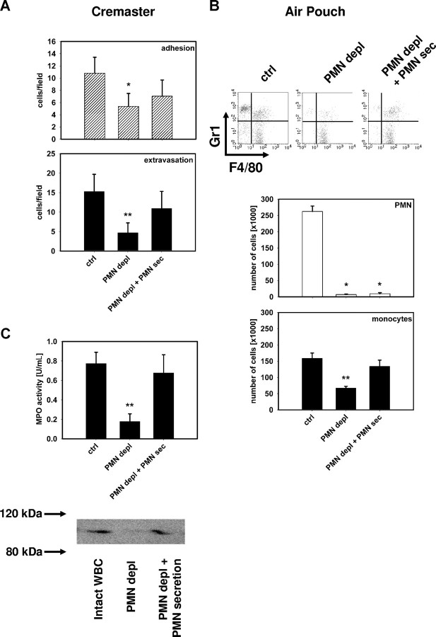
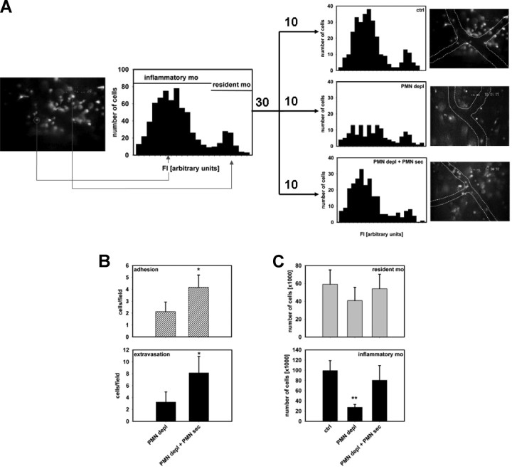
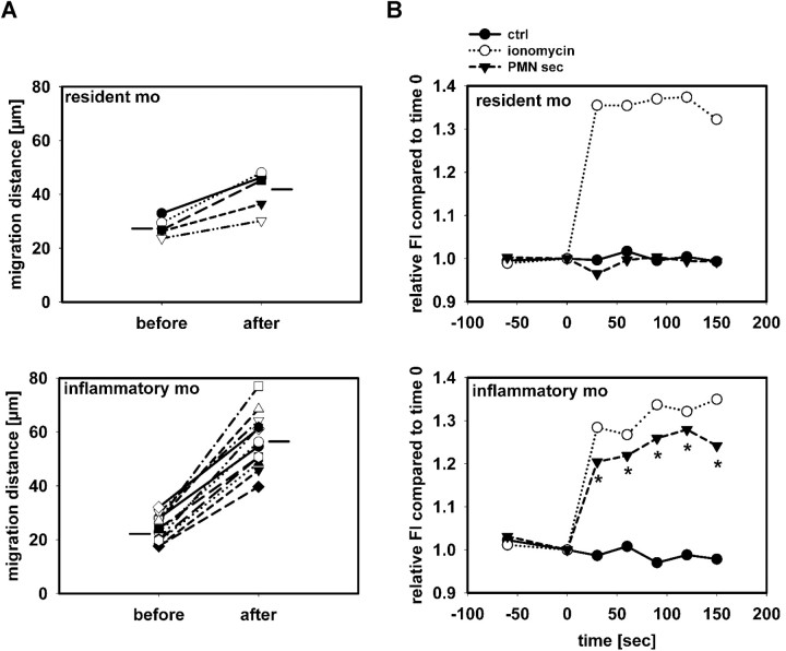
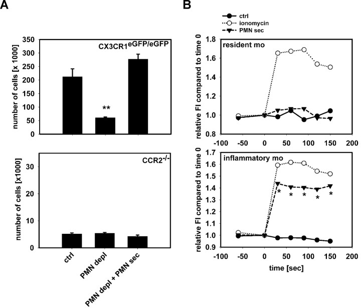
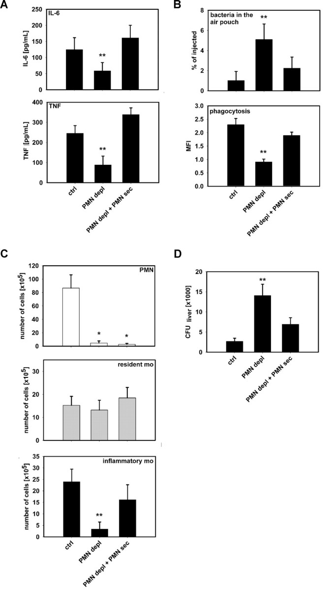
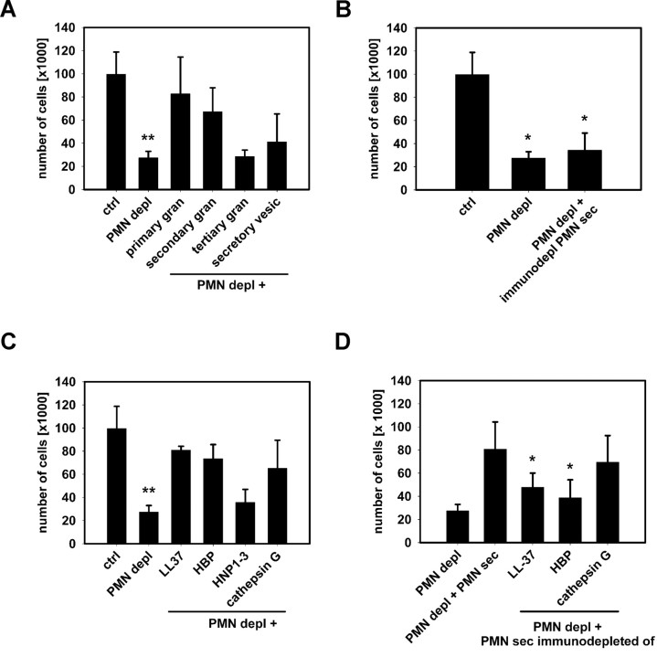
The leukocyte response in inflammation is characterized by an initial recruitment of polymorphonuclear leukocytes (PMN) preceding a second wave of monocytes to the site of injury or infection. In the mouse, 2 populations of monocytes have been identified, Gr1(-)CCR2(-)CX3CR1(hi) resident monocytes and Gr1(+)CCR2(+)CX3CR1(lo) inflammatory monocytes. Here, intravital microscopy of the musculus cremaster and a subcutaneous air pouch model were used to investigate a possible link between PMN extravasation and the subsequent emigration of inflammatory monocytes in response to local stimulation with PAF. In mice that were made neutropenic by injection of a PMN-depleting antibody, the extravasation of inflammatory monocytes, but not resident monocytes, was markedly reduced compared with mice with intact white blood cell count but was restored by local treatment with secretion of activated PMN. Components of the PMN secretion were found to directly activate inflammatory monocytes and further examination revealed PMN-derived LL-37 and heparin-binding protein (HBP/CAP37/azurocidin) as primary mediators of the recruitment of inflammatory monocytes via activation of formyl-peptide receptors. These data show that LL-37 and HBP specifically stimulate mobilization of inflammatory monocytes. This cellular cross-talk functionally results in enhanced cytokine levels and increased bacterial clearance, thus boosting the early immune response.


Figures

Figure 1
Figure 1
PMN secretion products mediate monocyte recruitment. (A) Number of adherent and extravasated fluorescent cells in the musculus cremaster quantified 12 hours after intrascrotal application of PAF (10−6 M) in CX3CR1eGFP/+ mice with intact WBC (ctrl), in neutropenic mice (PMN depl), and in neutropenic mice receiving intrascrotal injection of PMN secretion (PMN depl + PMN-sec). (B) Representative FACS plots showing Gr1 versus F4/80 staining and number of PMN and monocytes in the subcutaneous air pouch of C57BL/6 mice 12 hours after injection of PAF (10−6 M) in mice with intact WBC (ctrl), in neutropenic mice (PMN depl), and in neutropenic mice where PMN secretion was injected into the pouch (PMN depl + PMN-sec). (C) Analysis of MPO activity (top) and detection of MMP-9 (bottom) in the air pouch lavage fluid of C57BL/6 mice (ctrl), neutropenic mice (PMN depl), and neutropenic mice injected with PMN secretion (PMN depl + PMN-sec). Data are expressed as means (± SD), n = 4-6 for each bar. * indicates significant difference compared with ctrl; ** indicates significant difference compared both to ctrl and PMN depl + PMN-sec.
Figure 2
Figure 2
PMN secretion specifically mobilizes inflammatory monocytes. (A) Fluorescence intensity of extravasated monocytes in the musculus cremaster of CX3CR1eGFP/+ mice after intrascrotal injection of PAF (10−6 M). High-fluorescent cells represent the resident monocytes, whereas low-fluorescent cells constitute the inflammatory monocytes. Analyses were made in mice with intact WBC count (top right), in neutropenic mice (middle right), and in neutropenic mice treated intrascrotally with PMN secretion (bottom right). Data are based on analyses in 10 cremaster muscles in each group. Representative images are shown to the right of each histogram. (B) Adhesion and extravasation of inflammatory monocytes in the musculus cremaster 12 hours after intrascrotal injection of PAF (10−6 M). Analyses were made in neutropenic C57BL/6 mice with or without local treatment with PMN secretion. Ingestion of fluorescent latex beads allowed visualization of the inflammatory monocyte subset. * indicates significant difference compared with PMN depl. (C) Recruitment of resident and inflammatory monocytes in the air pouch of C57BL/6 mice 12 hours after stimulation with PAF (10−6 M). The monocytes were differentiated by FACS based on their Gr1 expression. Analyses were made in mice with intact WBC count (ctrl), neutropenic mice (PMN depl), and neutropenic mice treated with PMN secretion (PMN depl + PMN-sec). ** indicates significant difference compared with both ctrl and PMN depl + PMN-sec.
Figure 3
Figure 3
PMN secretion specifically activates inflammatory monocytes. (A) Migration distance of monocytes in 3 cremaster muscles of CX3CR1eGFP/+ mice over a 30-minute period before and after superfusion with PMN secretion. Distinction between resident and inflammatory monocytes was based on their fluorescence intensity. Horizontal lines indicate group average. The difference in number of cells included in the analysis reflects the different efficacy in recruitment between the 2 monocyte subsets. (B) Leukocytes from C57BL/6 mice were harvested by cardiac puncture, and intracellular Ca2+ mobilization was measured in resident monocytes (Gr1, F4/80+, top) and inflammatory monocytes (Gr1+, F4/80+, bottom) after stimulation with medium (ctrl), ionomycin, or PMN secretion (PMN-sec). Data were acquired before and at 30-second intervals after stimulation (time = 0) and presented as average of 4 to 6 analyses for each data point. * indicates significant difference between treatment with PMN secretion and control.
Figure 4
Figure 4
Recruitment of inflammatory monocytes by PMN secretion is CCR2-dependent. (A) Recruitment of Gr1+ inflammatory monocytes in the air pouch of CX3CR1eGFP/eGFP mice (top) and CCR2−/− (bottom) mice 12 hours after stimulation with PAF (10−6 M). Analyses were made in mice with intact WBC count (ctrl), neutropenic mice (PMN depl), and neutropenic mice treated with PMN secretion (PMN depl + PMN-sec). Note the difference in scale between the charts. ** indicates significant difference compared with both ctrl and PMN depl + PMN-sec. (B) Leukocytes from CCR2−/− mice were harvested by cardiac puncture and intracellular Ca2+ mobilization was measured in resident monocytes (Gr1, F4/80+, top) and inflammatory monocytes (Gr1+, F4/80+, bottom) after stimulation with medium (ctrl), ionomycin, or PMN secretion (PMN-sec). Data were acquired before and at 30-second intervals after stimulation (time = 0) and presented as average of 4 to 6 analyses for each data point. * indicates significant difference between treatment with PMN secretion and control.
Figure 5
Figure 5
PMN secretion stimulates cytokine release and bacterial phagocytosis by monocytes. (A) Concentrations of IL-6 (top) and TNF (bottom) in air pouch fluid of mice with intact WBC count (ctrl), neutropenic mice (PMN depl), and neutropenic mice treated with PMN secretion (PMN depl + PMN-sec). (B) Leukocyte recruitment in the air pouch was induced by PAF and fluorescent IgG-opsonized S aureus bacteria were injected 1 hour before the cells were harvested. Upper panel shows the number of bacteria retrieved from the air pouch fluid expressed in percentage of total amount of injected bacteria. Bottom panel shows fluorescence intensity of monocytes as a measure of bacterial phagocytosis. (C) Recruitment of PMN (top, Gr1+, F4/80), resident monocytes (middle, Gr1, F4/80+), and inflammatory monocytes (bottom, Gr1+, F4/80+) in the air pouch after injection of L monocytogenes (106). (D) Dissemination of L monocytogenes 24 hours after inoculation into the air pouch. Data show colony-forming units (CFU) in liver homogenates from mice with intact WBC, neutropenic mice, and neutropenic mice treated with PMN secretion. Data are expressed as means (± SD); n = 4-6 for each bar. * indicates significant difference compared with ctrl; ** indicates significant difference compared with both ctrl and PMN depl + PMN-sec.
Figure 6
Figure 6
Identification of HBP and LL-37 as principal mediators of PMN-induced mobilization of inflammatory monocytes. (A) Extravasation of Gr1+ inflammatory monocytes in the air pouch of neutropenic mice after stimulation with PAF and individual PMN granule fractions. ** indicates significant difference compared with both ctrl and PMN depl + primary or secondary granule fraction. (B) Immunodepletion of LL-37, HBP, HNP1-3, and cathepsin G from the PMN secretion, renders the secretion nonfunctional. * indicates significant difference compared with ctrl. (C) Extravasation of Gr1+ inflammatory monocytes in the air pouch of neutropenic mice after stimulation with PAF combined with recombinant/isolated forms of LL-37 (10 μg/mL), HBP (10 μg/mL), HNP1-3 (5 μg/mL), or cathepsin G (10 μg/mL). ** indicates significant difference compared with both ctrl and PMN depl + LL37/HBP/cathepsin G. (D) Extravasation of Gr1+ inflammatory monocytes in the air pouch of neutropenic mice after stimulation with PAF combined with crude PMN secretion or PMN secretion immunodepleted of LL-37, HBP, and cathepsin G, respectively. * indicates significant difference compared with PMN depl + PMN-sec. Data are expressed as means (± SD) n = 5 for each bar.
Figure 7
Figure 7
Involvement of FPRs in the PMN-induced mobilization of inflammatory monocytes. (A) Extravasation of Gr1+ inflammatory monocytes in the air pouch of neutropenic mice (PMN depl) after stimulation with PAF combined with PMN secretion or individual granule proteins. ▨ indicate pretreatment with the FPR antagonist BOC-PLPLP. * indicates significant difference compared with respective treatment in the absence of the fpr antagonist. (B) Ca2+ mobilization in inflammatory monocytes in response to PMN secretion or individual granule proteins. Ca2+ mobilization was measured in the FACS in the presence or absence of the FPR antagonist BOC-PLPLP. * indicates significant difference compared with respective treatment in the absence of the fpr antagonist.

References

    1. Witko-Sarsat V, Rieu P, Descamps-Latscha B, Lesavre P, Halbwachs-Mecarelli L. Neutrophils: molecules, functions and pathophysiological aspects. Lab Invest. 2000;80:617–653. - PubMed
    1. Doherty DE, Downey GP, Worthen GS, Haslett C, Henson PM. Monocyte retention and migration in pulmonary inflammation. Requirement for neutrophils. Lab Invest. 1988;59:200–213. - PubMed
    1. Janardhan KS, Sandhu SK, Singh B. Neutrophil depletion inhibits early and late monocyte/macrophage increase in lung inflammation. Front Biosci. 2006;11:1569–1576. - PubMed
    1. Fillion I, Ouellet N, Simard M, et al. Role of chemokines and formyl peptides in pneumococcal pneumonia-induced monocyte/macrophage recruitment. J Immunol. 2001;166:7353–7361. - PubMed
    1. Zhou J, Stohlman SA, Hinton DR, Marten NW. Neutrophils promote mononuclear cell infiltration during viral-induced encephalitis. J Immunol. 2003;170:3331–3336. - PubMed

Publication types