Review
. 2008 May;144(5):666-72.
doi: 10.1001/archderm.144.5.666.

Looking older: fibroblast collapse and therapeutic implications

Affiliations
Review

Looking older: fibroblast collapse and therapeutic implications

Gary J Fisher et al. Arch Dermatol. 2008 May.

Abstract

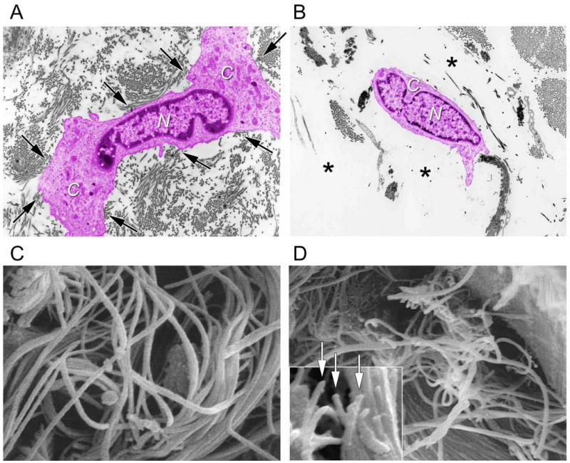
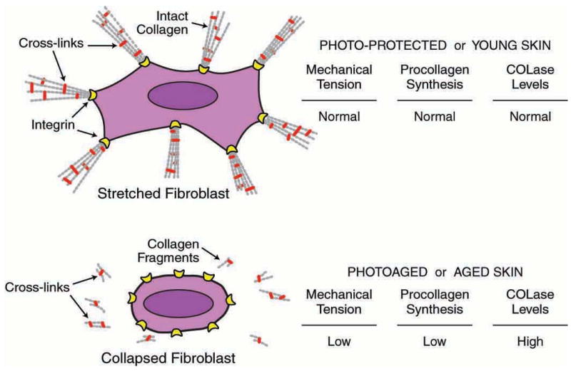
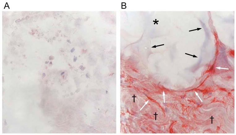
Skin appearance is a primary indicator of age. During the last decade, substantial progress has been made toward understanding underlying mechanisms of human skin aging. This understanding provides the basis for current use and new development of antiaging treatments. Our objective is to review present state-of-the-art knowledge pertaining to mechanisms involved in skin aging, with specific focus on the dermal collagen matrix. A major feature of aged skin is fragmentation of the dermal collagen matrix. Fragmentation results from actions of specific enzymes (matrix metalloproteinases) and impairs the structural integrity of the dermis. Fibroblasts that produce and organize the collagen matrix cannot attach to fragmented collagen. Loss of attachment prevents fibroblasts from receiving mechanical information from their support, and they collapse. Stretch is critical for normal balanced production of collagen and collagen-degrading enzymes. In aged skin, collapsed fibroblasts produce low levels of collagen and high levels of collagen-degrading enzymes. This imbalance advances the aging process in a self-perpetuating, never-ending deleterious cycle. Clinically proven antiaging treatments such as topical retinoic acid, carbon dioxide laser resurfacing, and intradermal injection of cross-linked hyaluronic acid stimulate production of new, undamaged collagen. Attachment of fibroblasts to this new collagen allows stretch, which in turn balances collagen production and degradation and thereby slows the aging process. Collagen fragmentation is responsible for loss of structural integrity and impairment of fibroblast function in aged human skin. Treatments that stimulate production of new, nonfragmented collagen should provide substantial improvement to the appearance and health of aged skin.


Conflict of interest statement

Conflict of Interest: Regents of the University of Michigan hold patents regarding the use of matrix metalloproteinase inhibitors for treatment and prevention of skin aging.

Figures

Figure 1
Figure 1
Fragmentation of collagen fibrils within dermis of aged/photoaged skin causes collapse of fibroblasts. A) Transmission electron micrograph of fibroblast (artificially colored pink for clarity) within dermis of young adult, sun-protected human skin. Note elongated, stretched appearance, and extended cytoplasm (C) away from nucleus (N) of fibroblast, which is in close proximity to abundant collagen fibrils (arrows, original magnification ×2,000). The top drawing of Figure 2 depicts a stretched fibroblast attached to intact collagen. B) Transmission electron micrograph of fibroblast (artificially colored for clarity) within dermis of photodamaged human skin. Note collapse of cytoplasm inward towards nucleus (N), and lack of adjacent collagen fibrils, compared to (A). Fibroblast is surrounded by amorphous material (*) The lower drawing of Figure 2 depicts a collapsed fibroblast surrounded by broken collgen. (C) Scanning electron micrograph of collagen fibrils in young adult human skin. Note that long fibrils are closely packed and fill space. Also note no apparent breaks in the fibrils (original magnification ×10,000). (D) Scanning electron micrograph (original magnification ×8,000) of collagen fibrils in photodamaged human skin. Note large gaps and numerous fragmented fibrils, compared to (C). Inset shows higher magnification of fragmented ends of fibrils (arrows, original magnification ×12,5000). Panels C and D are from Fligiel, S et al. Journal Investigative Dermatology, 120: 842-848, 2003.
Figure 2
Figure 2
Model depicting relationship between mechanical tension, and collagen production and fragmentation in human skin. In sun-protected young adult human skin, intact type I collagen fibrils in the dermis provide mechanical stability and attachment sites for fibroblasts. Receptors (integrins) on the surface of fibroblasts attach to collagen (and other proteins in the dermal extracellular matrix). Cytoskeletal machinery (actin-myosin microfilaments, not shown) within fibroblasts pulls on the intact collagen matrix, which in turn offers mechanical resistance. Dynamic mechanical tension that is created promotes assembly of intracellular scaffolding (microtubules/intermediate filaments, not shown), which pushes outward to cause fibroblasts to stretch. This stretch is required for fibroblasts to produce normal levels of collagen and MMPs. In photodamaged/aged human skin, attachments of fibroblasts to integrins are lost, and fragmented collagen fibrils fail to provide sufficient mechanical stability to maintain normal mechanical tension. Reduced mechanical tension causes fibroblasts to collapse, and collapsed fibroblasts produce less procollagen and more collagenase (COLase). Reduced collagen production and increased collagenase-catalyzed collagen fragmentation result in further reduction of mechanical tension, thereby causing continual loss of collagen.
Figure 3
Figure 3
Mechanical stretch induced by dermal injection of cross-linked hyaluronic acid filler stimulates collagen production in photodamaged human skin. (A) Saline vehicle (control) or (B) cross-linked hyaluronic acid filler (CLHA) was injected into photodamaged forearm skin. Skin biopsies were obtained four weeks post injection and analyzed for type I procollagen expression by immunohistochemistry. (A) Fibroblasts (nuclei stained blue) in saline-injected skin display barely-detectable procollagen expression (red stain). Also note amorphous space and fragmented thin appearance of collagen extracellular matrix (original magnification ×400). (B) In CLHA–injected skin, fibroblasts display intense type I procollagen immunostaining (red). CLHA appears as light blue material (black arrows) that occupies space adjacent to stretched fibroblasts (white arrows). Note also densely packed collagen fibrils (black daggers), not seen in saline-injected skin (A). These dense collagen fibrils are likely derived from CLHA-induced type I procollagen (i.e., conversion of type I procollagen into type I collagen) (original magnification ×400).

References

    1. Kligman AM. Early destructive effects of sunlight on human skin. JAMA. 1969;210:2377–2380. - PubMed
    1. Yaar M, Eller M, Gilchrest B. Fifty years of skin aging. J Invest Dermatol Sympos Proc. 2002;7:51–58. - PubMed
    1. Lavker R. Structural alterations in exposed and unexposed aged skin. J Invest Dermatol. 1979;73:559–566. - PubMed
    1. Scharffetter-Kochanek K. Skin aging. Clin Exp Dermatol. 2001;26(7):561. - PubMed
    1. Miyamura Y, Coelho SG, Wolber R, Miller SA, Wakamatsu K, Zmudzka BZ, Ito S, Smuda C, Passeron T, Choi W, Batzer J, Yamaguchi Y, Beer JZ, Hearing VJ. Regulation of human skin pigmentation and responses to ultraviolet radiation. Pigment Cell Res. 2007;20(1):2–13. - PubMed

Publication types