. 2008 Jun 3;105(22):7809-14.
doi: 10.1073/pnas.0711241105. Epub 2008 May 19.

Modulation of TNF-alpha-converting enzyme by the spike protein of SARS-CoV and ACE2 induces TNF-alpha production and facilitates viral entry

Affiliations

Modulation of TNF-alpha-converting enzyme by the spike protein of SARS-CoV and ACE2 induces TNF-alpha production and facilitates viral entry

Shiori Haga et al. Proc Natl Acad Sci U S A. .

Abstract

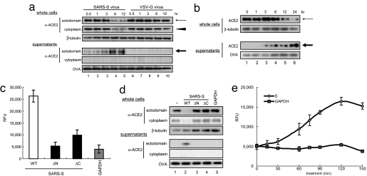
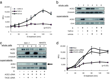
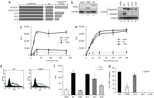
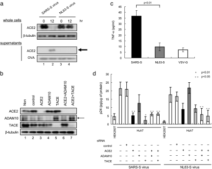
Severe acute respiratory syndrome coronavirus (SARS-CoV) is a high-risk infectious pathogen. In the proposed model of respiratory failure, SARS-CoV down-regulates its receptor, angiotensin-converting enzyme 2 (ACE2), but the mechanism involved is unknown. We found that the spike protein of SARS-CoV (SARS-S) induced TNF-alpha-converting enzyme (TACE)-dependent shedding of the ACE2 ectodomain. The modulation of TACE activity by SARS-S depended on the cytoplasmic domain of ACE2, because deletion mutants of ACE2 lacking the carboxyl-terminal region did not induce ACE2 shedding or TNF-alpha production. In contrast, the spike protein of HNL63-CoV (NL63-S), a CoV that uses ACE2 as a receptor and mainly induces the common cold, caused neither of these cellular responses. Intriguingly, viral infection, judged by real-time RT-PCR analysis of SARS-CoV mRNA expression, was significantly attenuated by deletion of the cytoplasmic tail of ACE2 or knock-down of TACE expression by siRNA. These data suggest that cellular signals triggered by the interaction of SARS-CoV with ACE2 are positively involved in viral entry but lead to tissue damage. These findings may lead to the development of anti-SARS-CoV agents.


Conflict of interest statement

The authors declare no conflict of interest.

Figures

Fig. 1.
Fig. 1.
Induction of ACE2 shedding by the specific binding of SARS-S and ACE2. (a) ACE2 shedding is induced by SARS-S. After infection with SARS-S, total extracts (Upper) or the supernatants (Lower) of cultured Vero E6 cells were analyzed. The thin arrow and arrowhead indicate the 120-kDa ACE2 protein detected by antibodies against the ectodomain and cytoplasmic domain, respectively. The bold arrow indicates the 80-kDa ACE2 detected by an antibody against the ACE2 cytoplasmic domain (lanes 2–5), which was not generated by control VSV-G virus (lanes 6–10). The proteins present in the culture supernatants were recovered with OVA by TCA precipitation. β-Tubulin was used as a loading control. (b) Recombinant SARS-S induces ACE2 shedding. Cells were treated with 100 μg/ml SARS-S (residues 284–541), and ACE2 in the culture supernatant was examined as described in a. (c) Binding of the SARS-S mutants to ACE2. Two SARS-S mutants, ΔN (residues 332–541) and ΔC (residues 284–490), were prepared according to a report showing the minimum region of SARS-S required for ACE2 binding (27), and their binding activities to ACE2 were examined (see also Materials and Methods). (d) Specific binding to ACE2 is required for ACE2 shedding. After treatment with the ΔN and ΔC SARS-Ss, the amount of ACE2 in the culture supernatant was examined as described in a. (e) C-peptidase activity of the shed ACE2. The C-peptidase activity in the culture supernatant of SARS-S-treated Vero E6 cells was measured (filled diamonds) (see Materials and Methods). GAPDH was used as a negative control (filled squares).
Fig. 2.
Fig. 2.
ACE2 shedding depends on TACE. (a) Inhibitory effects of TAPI-0 on ACE2 shedding. Vero E6 cells were cultured with or without 100 nM TAPI-0, and the amounts of C-peptidase activity in the culture supernatants were measured. SARS-S was used at a concentration of 100 μg/ml. (b) TACE inhibitor blocked SARS-S-induced ACE2 shedding. The experiments described in a were carried out and ACE2 was detected by Western blot analysis. (c) ACE2 shedding depends on TACE activity. tace-KO cells were transfected with cDNAs encoding ACE2 or TACE and then treated with 100 μg/ml SARS-S for 6 h. Shed ACE2 was then detected (bold arrow). (d) The effects of TACE siRNA on ACE2 shedding. After the introduction of TACE or control siRNA, SARS-S-induced ACE2 shedding was examined by measuring the amount of C-peptidase activity in the culture supernatant. Huh-7 cells, a human cell line that expresses endogenous ACE2 and TACE (see Fig. 5c), were analyzed.
Fig. 3.
Fig. 3.
ACE2 shedding by SARS-S requires the cytoplasmic domain of ACE2. (a) Schematic diagrams of the ACE2 cytoplasmic domain mutants. ΔTMC-ACE2 contains only the ectodomain of ACE2 with the transmembrane domain of PDGF receptor. (b) Expression of the ACE2 cytoplasmic domain mutants. The molecular weights of full-length ACE2 (lane 3) and all mutants except ΔTMC-ACE2 (lanes 6–8) were ≈120 kDa. The molecular weight of ΔTMC-ACE2 was ≈95 kDa (lane 2). (c) ΔTMC-ACE2 did not show ectodomain shedding with SARS-S. After treatment with SARS-S, the amounts of C-peptidase activity in the culture supernatant of cells expressing WT (filled sqaures) or ΔTMC-ACE2 (filled diamonds) were examined. (d) Binding of ΔTMC-ACE2 to SARS-S. Recombinant SARS-S was expressed and purified as a chimeric protein with the Fc portion of human IgG (Fc), and bound SARS-S was detected by FACS analysis, using FITC-labeled anti-human IgG (9). Each line indicates the cells that were positive for bound SARS-S. Solid peaks depict cells bound nonspecifically with control IgG-Fc. (e) ΔTMC-ACE2 was competent for ectodomain shedding by PMA. After treatment with PMA for 1 h, the amounts of C-peptidase activity in the culture supernatant of cells expressing WT (filled squares) or ΔTMC-ACE2 (filled diamonds) were examined. (f) The ectodomain shedding of ACE2 depends on the cytoplasmic domain. The levels of C-peptidase activity in the culture supernatant of cells expressing various ACE2 mutants were measured. (g) TACE activation requires the cytoplasmic domain of ACE2. TACE activation, judged by the production of TNF-α in the culture supernatant, was induced by both WT and ΔC17-ACE2. TNF-α was detected 20 h after SARS-S treatment.
Fig. 4.
Fig. 4.
The cytoplasmic tail of ACE2 and TACE promote infection by SARS-CoV. (a) No difference in infection efficiency was detected by reporter assay, using a pseudotyped lentivirus system. After infection of HEK293T cells expressing WT or ΔTMC-ACE2 with a pseudotyped lentivirus encoding luciferase, luciferase activity was measured. Luciferase activity was assayed in triplicate. (b) Detection of differences in viral infection efficiency based on the intracellular p24 level. The experiment in a was followed by measurement of the intracellular p24 level, using an ELISA kit. (c) A marked difference in viral infection efficiency detected by real-time RT-PCR. Each cell was harvested and analyzed 2 h after infection with SARS-CoV. The data were normalized by 18S ribosomal RNA. (d) The cytoplasmic tail is required for efficient viral infection. SARS-CoV was used to infect cells expressing deletion mutants of the cytoplasmic tail of ACE2 (Fig. 3a). Then, real-time RT-PCR was carried out as described in c. (e) TACE is required for efficient viral entry. The efficiency of viral entry into Huh-7 cells was examined before and after the introduction of TACE or ACE2 siRNAs. TACE siRNA [but not ADAM10 (metalloprotease control) siRNA] significantly attenuated viral entry (P < 0.05). ACE2 siRNA also decreased viral infection (P < 0.01).
Fig. 5.
Fig. 5.
NL63-S does not induce ACE2 shedding. (a) No ACE2 shedding was observed when using NL-63-S. After infection of Vero E6 cells with NL63-S or SARS-S, ACE2 was examined as described in Fig. 1a. (b) NL63-S does not induce TNF-α production. TNF-α was measured in the culture supernatants of ACE2-expressing HEK293T cells. The supernatants were collected 12 h after infection with NL63-S or SARS-S. (c) Effects of siRNAs on the expression of endogenous gene products. Huh-7 cells were transduced with siRNAs against TACE, ACE2, and ADAM10, and the expression of each protein was examined. Lanes 3, 4, and 5 indicate the endogenous expression of ACE2, ADAM10, and TACE, respectively. Lanes 6 and 7 depict the effects of the combined use of siRNAs on protein expression. (d) The differential requirements of TACE for viral entry by NL63-S and SARS-S. Huh-7 cells were infected with SARS-S and NL63-S viruses for 4 h, and the efficiency of viral entry was examined by measuring the intracellular p24 level. Note that the entry of SARS-S was reduced by siRNAs against ACE2 and TACE, whereas NL63-S was not.

Similar articles

Cited by

References

    1. Kaiazek TG, et al. A novel coronavirus associated with severe acute respiratory syndrome. N Engl J Med. 2003;15:1953–1966. - PubMed
    1. Tipnis SR, et al. A human homolog of angiotensin-converting enzyme. Cloning and functional expression as a captopril-insensitive carboxypeptidase. J Biol Chem. 2000;275:33238–33243. - PubMed
    1. Nicholls J, Peiris M. Good ACE, bad ACE do battle in lung injury, SARS. Nat Med. 2005;11:821–822. - PMC - PubMed
    1. Li W, et al. Angiotensin-converting enzyme 2 is a functional receptor for the SARS coronavirus. Nature. 2003;426:450–454. - PMC - PubMed
    1. Kuba K, et al. A crucial role of angiotensin converting enzyme 2 (ACE2) in SARS coronavirus-induced lung injury. Nat Med. 2005;11:875–879. - PMC - PubMed

Publication types

MeSH terms