. 2008 May 27;105(21):7534-9.
doi: 10.1073/pnas.0802865105. Epub 2008 May 19.

Luteolin reduces IL-6 production in microglia by inhibiting JNK phosphorylation and activation of AP-1

Affiliations

Luteolin reduces IL-6 production in microglia by inhibiting JNK phosphorylation and activation of AP-1

Saebyeol Jang et al. Proc Natl Acad Sci U S A. .

Abstract

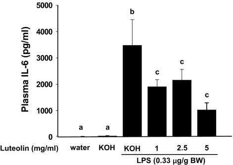
Luteolin, a flavonoid found in high concentrations in celery and green pepper, has been shown to reduce production of proinflammatory mediators in LPS-stimulated macrophages, fibroblasts, and intestinal epithelial cells. Because excessive production of proinflammatory cytokines by activated brain microglia can cause behavioral pathology and neurodegeneration, we sought to determine whether luteolin also regulates microglial cell production of a prototypic inflammatory cytokine, IL-6. Pretreatment of primary murine microlgia and BV-2 microglial cells with luteolin inhibited LPS-stimulated IL-6 production at both the mRNA and protein levels. To determine how luteolin inhibited IL-6 production in microglia, EMSAs were performed to establish the effects of luteolin on LPS-induced binding of transcription factors to the NF-kappaB and activator protein-1 (AP-1) sites on the IL-6 promoter. Whereas luteolin had no effect on the LPS-induced increase in NF-kappaB DNA binding activity, it markedly reduced AP-1 transcription factor binding activity. Consistent with this finding, luteolin did not inhibit LPS-induced degradation of IkappaB-alpha but inhibited JNK phosphorylation. To determine whether luteolin might have similar effects in vivo, mice were provided drinking water supplemented with luteolin for 21 days and then they were injected i.p. with LPS. Luteolin consumption reduced LPS-induced IL-6 in plasma 4 h after injection. Furthermore, luteolin decreased the induction of IL-6 mRNA by LPS in hippocampus but not in the cortex or cerebellum. Taken together, these data suggest luteolin inhibits LPS-induced IL-6 production in the brain by inhibiting the JNK signaling pathway and activation of AP-1 in microglia. Thus, luteolin may be useful for mitigating neuroinflammation.


Conflict of interest statement

The authors declare no conflict of interest.

Figures

Fig. 1.
Fig. 1.
Suppression of LPS-induced IL-6 secretion in primary microglia and BV-2 cells. Primary microglia (A) and BV-2 cells (B) were pretreated with luteolin for 1 h and stimulated with LPS for a 24 h incubation period. IL-6 secretion was measured by ELISA. Concentration of IL-6 in supernatants from primary microglia and BV-2 cells stimulated with LPS was 1,100 and 2,800 pg/ml, respectively. Data are presented as percentage of IL-6 release compared with IL-6 level of LPS alone treatment (100%). Bars represent the mean ± SEM from three independent experiments. Means with different letters are different (P < 0.05).
Fig. 2.
Fig. 2.
Suppression of LPS-induced IL-6 mRNA expression in primary microglia and BV-2 cells. Primary microglia (A) and BV-2 cells (B) were pretreated with luteolin for 1 h and stimulated with LPS for an 8-h incubation period. Expression of IL-6 mRNA was measured by RT-PCR. In primary microglia the threshold cycle of GAPDH and IL-6 was 32 and 19, respectively. In BV-2 cells the threshold cycle of GAPDH and IL-6 was 27 and 13, respectively. Data are presented as percentage of IL-6 mRNA expression compared with IL-6 mRNA expression level of LPS-alone treatment (100%). Bars represent the mean ± SEM from three independent experiments. Means with different letters are different (P < 0.05).
Fig. 3.
Fig. 3.
Luteolin inhibits LPS-induced AP-1 activation. BV-2 cells were pretreated with luteolin for 1 h and stimulated with LPS for a 1 h incubation period. (A and B) Nuclei were extracted for NF-κB (A) and AP-1 (B) EMSA. PDTC was used as a NF-κB inhibitor. (C and D) Transcription factor binding activity for NF-κB (C) and AP-1 (D) was determined by densitometric scanning. Bars represent the mean ± SEM from three independent experiments. Means with different letters are different (P < 0.05).
Fig. 4.
Fig. 4.
Luteolin inhibits LPS-induced phosphorylation of JNK. BV-2 cells were pretreated with luteolin for 1 h and stimulated with LPS for a 30-min incubation period. Cells were collected, and cell lysates were separated by 12% SDS/PAGE, transferred to membranes, and blotted with specific antibodies to IκB-α (A), Erk and phosphorylated Erk (B), p38 and phosphorylated p38 (C), and JNK and phosphorylated JNK (D). β-Actin was blotted as an internal control. Bars represent the mean ± SEM from three independent experiments. Means with different letters are different (P < 0.05).
Fig. 5.
Fig. 5.
Luteolin reduces LPS-stimulated IL-6 production in plasma. Mice fed luteolin (0, 1, 2.5, or 5 mg/ml) for 21 days were injected with LPS (0.33 μg/g BW). At 4 h postinjection, mice were killed, and plasma was collected for IL-6 measurement. Bars represent the mean ± SEM (n = 9). Means with different letters are different (P < 0.05).
Fig. 6.
Fig. 6.
Luteolin reduces LPS-stimulated IL-6 production in hippocampus. Mice fed luteolin (0, 1, 2.5, or 5 mg/ml) for 21 days were injected with LPS (0.33 μg/g BW). Four hours after LPS injection, cortex (Left), cerebellum (Center), and hippocampal (Right) tissue were collected, and IL-6 mRNA was measured by RT-PCR. Bars represent the mean ± SEM (n = 9). Means with different letters are different (P < 0.05).

Similar articles

Cited by

References

    1. Laflamme N, Rivest S. Toll-like receptor 4: The missing link of the cerebral innate immune response triggered by circulating gram-negative bacterial cell wall components. FASEB J. 2001;15:155–163. - PubMed
    1. Guha M, Mackman N. LPS induction of gene expression in human monocytes. Cell Signal. 2001;13:85–94. - PubMed
    1. Dantzer R, Kelley KW. Twenty years of research on cytokine-induced sickness behavior. Brain Behav Immun. 2007;21:153–160. - PMC - PubMed
    1. Godbout JP, et al. Exaggerated neuroinflammation and sickness behavior in aged mice following activation of the peripheral innate immune system. FASEB J. 2005;19:1329–1331. - PubMed
    1. Godbout JP, et al. Aging exacerbates depressive-like behavior in mice in response to activation of the peripheral innate immune system. Neuropsychopharmacology. 2007 in press. - PMC - PubMed

Publication types

MeSH terms

LinkOut - more resources