. 2008 Jun 1;180(11):7385-93.
doi: 10.4049/jimmunol.180.11.7385.

CD43 regulates Th2 differentiation and inflammation

Affiliations

CD43 regulates Th2 differentiation and inflammation

Judy L Cannon et al. J Immunol. .

Abstract

CD43 is a highly glycosylated transmembrane protein that regulates T cell activation. CD43(-/-) T cells are hyperproliferative and the cytoplasmic tail of CD43 has been found to be sufficient to reconstitute wild-type proliferation levels, suggesting an intracellular mechanism. In this study, we report that upon TCR ligation CD43(-/-) T cells demonstrated no increase in tyrosine phosphorylation but a decreased calcium flux. Interestingly, CD43(-/-) T cells preferentially differentiated into Th2 cells in vitro, and CD43(-/-) T cells show increased GATA-3 translocation into the nucleus. In vivo, CD43(-/-) mice exhibited increased inflammation in two separate models of Th2-mediated allergic airway disease. In contrast, in Th1-mediated diabetes, nonobese diabetic CD43(-/-) mice did not significantly differ from wild-type mice in disease onset or progression. Th1-induced experimental autoimmune encephalomyelitis to MOG(35-55) was also normal in the CD43(-/-) mice. Nonetheless, the CD43(-/-) mice produced more IL-5 when restimulated with MOG(35-55) in vitro and demonstrated decreased delayed-type hypersensitivity responses. Together, these data demonstrate that although CD43(-/-) T cells preferentially differentiate into Th2 cells, this response is not sufficient to protect against Th1-mediated autoimmune responses.


Conflict of interest statement

Disclosures: The authors have no financial conflict of interest.

Figures

FIGURE 1
FIGURE 1
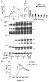
CD43−/− T cells are hyperproliferative, yet have a decreased Ca2+ flux. A, DO.11.10 and DO.CD43−/− T cells were cultured with A20 cells and 0.03 μg/ml OVA peptide. Plates were pulsed with 1 μCi/well [3H]thymidine 8–12 h before being harvested. B, DO.11.10 and DO.CD43−/− T cells were incubated with CFSE, then washed and cultured with A20 cells and 0.03 μg/ml OVA peptide. On day 3, the cells were harvested, stained with KJ1-26, and CFSE intensity was analyzed by flow cytometry. Data presented in A and B are representative of >10 independent experiments. C, BALB.CD43+/− and BALB.CD43−/− T cells were activated by cross-linking the TCR for indicated times. Lysates were precipitated with anti-P-Tyr (FB2), and immunoblotted with anti-P-Tyr (4G10). Data shown are representative of four independent experiments. D, Fura Red- and Fluo 3-dyed B6 and B6.CD43−/− T cells were incubated with 5 μg/ml anti-CD3 for 10 min at room temperature immediately before acquisition. A control sample was run for 1 min before being replaced with anti-CD3-incubated sample for 1 min (first arrow). Goat anti-hamster cross-linker was added 2 min into acquisition (second arrow). Data shown are representative of three independent experiments.
FIGURE 2
FIGURE 2
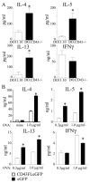
CD43−/− T cells produce increased amounts of IL-4, IL-5, and IL-13 and reintroduction of CD43 into CD43−/− T cells reconstitutes WT levels of cytokine production. A, DO.11.10 and DO.CD43−/− T cells were cultured with irradiated splenocytes and 1 μg/ml OVA323–339 peptide for 7 days, then restimulated for 48 h on anti-CD3-coated plates before assaying for IL-4, IL-5, IL-13, and IFN-γ by ELISA. (All * are p < 0.05.) Data are representative of four independent experiments. B, DO.CD43−/− T cells were transduced with either CD43FLeGFP or a control eGFP vector, sorted for GFP+ cells, and restimulated with irradiated splenocytes and indicated concentrations of OVA323–339 peptide for 48 h. Transduction efficiency was between 20 and 40% and, after sorting, >98% GFP+. Super-natants were taken for cytokine analysis. (All * are p < 0.0001, except IL-4, p = 0.0015 and IFN-γ, p = 0.0002).
FIGURE 3
FIGURE 3
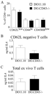
Memory CD43−/− T cells do not produce more early IL-4. A, Total ex vivo lymph node cells from DO.11.10 and DO.CD43−/− mice were stained for CD4 and either CD45RB, CD44, or CD62L. B, DO.11.10 and DO.CD43−/− T cells were cultured with irradiated APCs and 1 μg/ml OVA323–339 peptide for 48 h, then assayed for IL-4. Data are representative of five independent experiments.
FIGURE 4
FIGURE 4
CD43−/− T cells show greater GATA-3 nuclear translocation than WT T cells. DO11.10 and DOCD43−/− T cells were purified, stimulated with irradiated splenocytes and 0.1 μg/ml OVA peptide with and without 2 ng/ml IL-4 for 3 days, harvested, lysed, and nuclear extracts were prepared and analyzed by SDS-PAGE and blotted for GATA-3 and lamin A. Quantification was performed by calculating [GATA-3]/[lamin A] and normalizing to the WT condition.
FIGURE 5
FIGURE 5
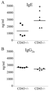
Unimmunized BALB/c.CD43−/− mice have increased levels of serum IgE. Sera from BALB/c.CD43+/− (n = 5) and BALB/c.CD43−/− (n = 5) littermates were collected and IgE (A) and IgG2a (B) levels were assayed. (IgE, p < 0.05).
FIGURE 6
FIGURE 6
CD43−/− mice have increased Th2-mediated airway inflammation. A, BALB/c (n = 9) and BALB/c.CD43−/− (n = 12) mice were sensitized and challenged with OVA peptide, and total number of eosinophils in the BAL was determined by differential counts (p < 0.01). B, Lungs from the OVA- sensitized and challenged mice in A were prepared for histological analysis and scored for inflammation severity around the vessels and bronchioles on a scale of 0–5 (All * are p < 0.005). C, B6 (n = 10) and B6.CD43−/− (n = 9) mice were sensitized with S. mansoni eggs and challenged with SEA. Total number of eosinophils, T cells, and macrophages in the BAL were determined by flow cytometry (eosinophils, p < 0.005). D, Total lung extracts from SCH mice were tested for cytokine content (IL-5, p < 0.005). E, Sera from age-matched B6 (n = 9) and B6.CD43−/− (n = 9) mice were collected before and after sensitization and challenge with S. mansoni eggs and SEA and analyzed for IgE. Before SCH, B6.CD43−/− mice had significantly more serum IgE than B6 (p < 0.05). This difference was also seen between B6 and B6.CD43−/− following SCH (p < 0.0001) and, as expected, IgE serum levels increased after SCH within each strain (p < 0.0001). F and G, Representative H&E-stained histological sections from lungs of WT (F) or CD43−/− (G) mice sensitized and challenged with SEA.
FIGURE 7
FIGURE 7
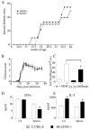
CD43−/− mice have normal incidence and progression of diabetes and EAE, but increased IL-5 and decreased DTH responses to MOG35–55. A, NOD.CD43+/− and NOD.CD43−/− mice were tested weekly for blood glucose levels. Mice were considered diabetic when glucose levels went above 250 mg/dl. B, B6 and B6.CD43−/− mice were treated with M. tuberculosis and MOG35–55 and evaluated for clinical symptoms of EAE on a scale of 0–5 as described in Materials and Methods. C, B6 and B6.CD43−/− were injected with no (−) or 10 μg (+) of MOG35–55 and ear swelling was measured 24 h later (p < 0.02). Lymph node and spleen cells taken on day 10 from B6 and B6.CD43−/− mice that had been primed with MOG35–55/CFA and cultured with 25 μM MOG35–55 for 48 h were assayed for IFN-γ (D) and IL-5 (E). IFN-γ, Spleen, p < 0.02; IL-5, lymph node (LN), p < 0.01; Spleen, p < 0.01.

References

    1. O'Garra A, Arai N. The molecular basis of T helper 1 and T helper 2 cell differentiation. Trends Cell Biol. 2000;10:542–550. - PubMed
    1. Constant SL, Bottomly K. Induction of Th1 and Th2 CD4+ T cell responses: the alternative approaches. Annu Rev Immunol. 1997;15:297–322. - PubMed
    1. Boutin Y, Leitenberg D, Tao X, Bottomly K. Distinct biochemical signals characterize agonist- and altered peptide ligand-induced differentiation of naive CD4+ T cells into Th1 and Th2 subsets. J Immunol. 1997;159:5802–5809. - PubMed
    1. Rulifson IC, Sperling AI, Fields PE, Fitch FW, Bluestone JA. CD28 costimulation promotes the production of Th2 cytokines. J Immunol. 1997;158:658–665. - PubMed
    1. Dong C, Juedes AE, Temann UA, Shresta S, Allison JP, Ruddle NH, Flavell RA. ICOS co-stimulatory receptor is essential for T-cell activation and function. Nature. 2001;409:97–101. - PubMed

Publication types

MeSH terms