. 2008 Jun 1;180(11):7565-73.
doi: 10.4049/jimmunol.180.11.7565.

Mast cell cathelicidin antimicrobial peptide prevents invasive group A Streptococcus infection of the skin

Affiliations

Mast cell cathelicidin antimicrobial peptide prevents invasive group A Streptococcus infection of the skin

Anna Di Nardo et al. J Immunol. .

Abstract

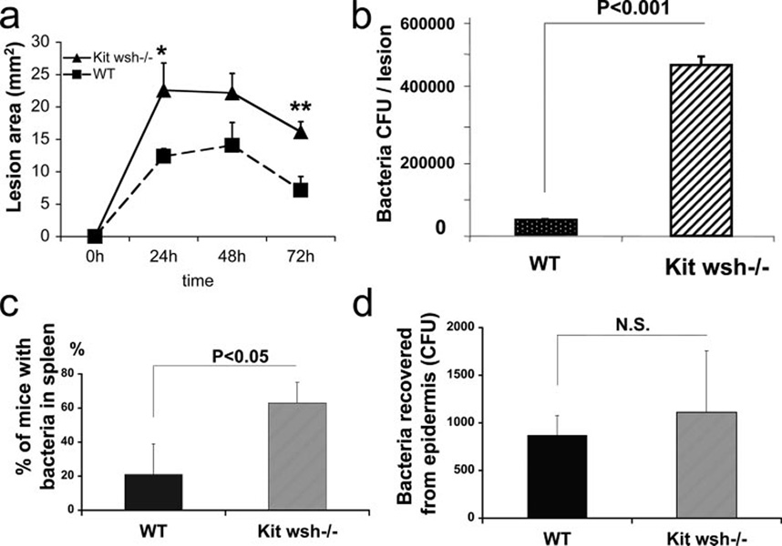
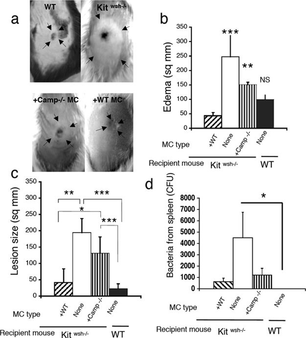
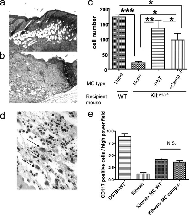
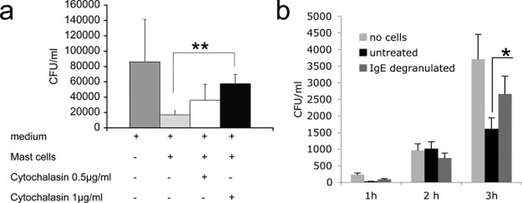
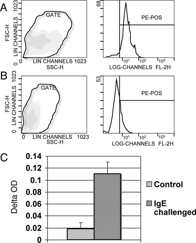
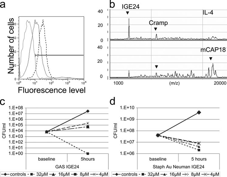
Mast cells (MC) express cathelicidin antimicrobial peptides that act as broad-spectrum antibiotics and influence the immune defense of multiple epithelial surfaces. We hypothesized that MC help protect against skin infection through the expression of cathelicidin. The susceptibility of MC-deficient mice (Kit Wsh(-/-)) to invasive group A streptococcus (GAS) was compared with control mice. Following s.c. injection of GAS, MC-deficient mice had 30% larger skin lesions, 80% more lesional bacteria, and 30% more spleens positive for bacteria. In contrast to results obtained when GAS was injected into skin, no significant differences were noted between MC-deficient mice and control mice after GAS was applied topically, indicating that MC activity is most important after barrier penetration. To determine whether these differences were due to MC expression of cathelicidin, MC-deficient mice were reconstituted with MC derived from either wild-type or cathelicidin-deficient (Camp(-/-)) mice and challenged with GAS. Forty-eight hours after bacterial injection, mice that did not receive MC had an average lesion size of 200 mm(2), mice reconstituted with wild-type MC showed lesions comparable to control mice (25 mm(2)), while mice reconstituted with Camp(-/-) MC showed an average lesion size of 120 mm(2). Surface-enhanced laser desorption/ionization time-of-flight mass spectrometry (SELDI-TOF-MS) analysis of cathelicidin peptide purified from mast cells defined this as a unique 28-aa peptide. Combined, these results show that MC confer defense against Gram-positive bacterial infection in the skin, a function mediated in part by the expression of a unique cathelicidin peptide.


Conflict of interest statement

Disclosures

The authors have no financial conflict of interest.

Figures

FIGURE 1
FIGURE 1
MC-deficient mice are more susceptible to skin infection and develop bacteremia. a, Wound area (mm2) over time in WT and MC-deficient mice infected with GAS.*, p < 0.05 and **, p < 0.01 b, Enumeration of GAS recovered from excised lesions of mice sacrificed on day 4 following s.c. infection, p < 0.001. c, Percentage of mice containing bacteria in the spleen, p < 0.05. d, Enumeration of GAS recovered from the superficial layer of the epidermis following topical infection.
FIGURE 2
FIGURE 2
MC-deficient mice (Kit Wsh−/−) reconstituted with WT and Camp−/− MC in the skin. a, Representative lesions from MC-deficient mice (Kit Wsh−/−), MC-deficient mice (Kit Wsh−/−) reconstituted with Camp−/− (Kit Wsh−/− plus Camp−/− MC) MC, MC-deficient mice (Kit Wsh−/−) reconstituted with WT MC (Kit Wsh−/− plus WT MC), and WT mice at 24 h following GAS infection. Arrows indicate the margins of edema visible on the skin surface. b, Measurement of visible edema on skin (mm2) 24 h after GAS infection in WT, MC-deficient mice (Kit Wsh−/−), and MC-deficient mice receiving adoptive transfer of WT MC (Kit Wsh−/− +WT MC) or MC derived from cathelicidin-deficient mice (+Camp−/− MC). c, Measurement of GAS-erosive lesion size (mm2) 48 h after bacterial injection in WT mice, MC-deficient mice (Kit Wsh−/−), and MC-deficient mice receiving adoptive transfer of WT MC (Kit Wsh−/−plus WT MC) or MC derived from cathelicidin-deficient mice (+Camp−/− MC). d, Number of bacteria in spleens containing bacteria at 72 h postinfection in WT mice and MC-deficient mice (Kit Wsh−/−) receiving adoptive transfer of WT MC (+WT), no transplant (none) or MC derived from cathelicidin-deficient mice (+Camp−/−), *, p < 0.05.
FIGURE 3
FIGURE 3
The presence of MC during infection modifies neutrophil recruitment in the upper dermis. a, H&E staining of skin in an area adjacent to the infected lesion in a WT mouse. b, H&E staining of skin in an area adjacent to the infected lesion in MC-deficient mice (Kit Wsh−/−). c, Measurement of the neutrophil infiltrate in the upper dermis at 72 h. Cell infiltrate was manually counted in three adjacent sections next to the site of infection. Data shown are cell number observed in WT mice, MC-deficient mice (Kit Wsh−/−) without transplant (none) and MC-deficient mice receiving adoptive transfer of WT MC (+WT) or MC derived from cathelicidin-deficient mice (+Camp−/−), *, p < 0.05; **, p < 0.01; and ***, p < 0.001. d, CD117 immunoperoxidase staining of skin in an area adjacent to the infected lesion in MC-deficient mice (Kit Wsh−/−) reconstituted with MC derived from cathelicidin-deficient mice. The arrows indicate positive cells. e, Measurement of the CD117-positive infiltrate in the dermis at 72 h. Cell infiltrate was manually counted in three adjacent sections next to the site of infection. Data shown are cell number observed in WT mice, MC-deficient mice (Kit Wsh−/−) without transplant, and MC-deficient mice receiving adoptive transfer of WT MC (+WT) or MC derived from cathelicidin-deficient mice (+Camp−/−).
FIGURE 4
FIGURE 4
MC use cathelicidin to kill GAS intracellularly. a, Enumeration of GAS following incubation with MC treated with or without cytochalasin for 3 h. *, p < 0.05 and **, p < 0.01. b, Enumeration of GAS incubated with degranulated MC, *, p < 0.05.
FIGURE 5
FIGURE 5
MC degranulation assay and cathelicidin release. MC degranulation was confirmed by two different systems. FACS analysis of side scatter show cells granularity before (left a) and after degranulation (left b). Before degranulation, cells are bigger and present higher side scatter level. Cathelicidin content was measured simultaneously in the same cell populations with FITC anti-Cramp (right a and b). c, After degranulation, medium was analyzed to confirm degranulation by d-β-glucosaminidase assay; results were read with a spectrophotometer and plotted as ΔOD.
FIGURE 6
FIGURE 6
Stimulated MC increase cathelicidin expression and process cathelicidin to smaller peptides. a, FACS analysis of cathelicidin expression in MC stimulated with IL-4 (10 ng/ml). The dotted line represents IL-4-treated cells, the gray line the untreated, and the black the IgG-negative control. b, Identification of a novel processed form of MC cathelicidin, IGE24, by SELDI-TOF-MS. MC were stimulated with IL-4, harvested with acetic acid, lyophilized, resuspended in MOPS buffer, and analyzed by SELDI-TOF using a polyclonal cathelicidin Ab. Mass of peptide indicated by arrow is 2805 kDa, corresponding to sequence IGEKLKKI GQKI KNFFQKLVPQ PEQ. In the control lane of the SELDI-TOF, peaks are visible corresponding to the CRAMP peptide and to the full-length peptide mCAP18. c, Minimal inhibitory concentration of synthetic IGE24 peptide against GAS following 5-h incubation in RPMI 1640/10% FCS. d, Minimal inhibitory concentration of synthetic IGE24 peptide against S. aureus following 5-h incubation in RPMI 1640/10% FCS.

References

    1. Malaviya R, Ikeda T, Ross EA, Jakschik BA, Abraham SN. Bacteria: mast cell interactions in inflammatory disease. Am. J. Ther. 1995;2:787–792. - PubMed
    1. Malaviya R, Abraham SN. Mast cell modulation of immune responses to bacteria. Immunol. Rev. 2001;179:16–24. - PubMed
    1. Di Nardo A, Vitiello A, Gallo RL. Cutting edge: mast cell antimicrobial activity is mediated by expression of cathelicidin antimicrobial peptide. J. Immunol. 2003;170:2274–2278. - PubMed
    1. Nizet V, Ohtake T, Lauth X, Trowbridge J, Rudisill J, Dorschner RA, Pestonjamasp V, Piraino J, Huttner K, Gallo RL. Innate antimicrobial peptide protects the skin from invasive bacterial infection. Nature. 2001;414:454–457. - PubMed
    1. Skerlavaj B, Scocchi M, Gennaro R, Risso A, Zanetti M. Structural and functional analysis of horse cathelicidin peptides. Antimicrob. Agents Chemother. 2001;45:715–722. - PMC - PubMed

Publication types

MeSH terms

Substances