. 2008 Sep 11;27(40):5288-302.
doi: 10.1038/onc.2008.161. Epub 2008 May 19.

Roles of cyclins A and E in induction of centrosome amplification in p53-compromised cells

Affiliations

Roles of cyclins A and E in induction of centrosome amplification in p53-compromised cells

K Hanashiro et al. Oncogene. .

Abstract

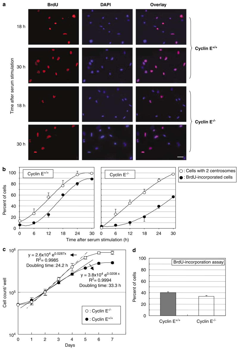
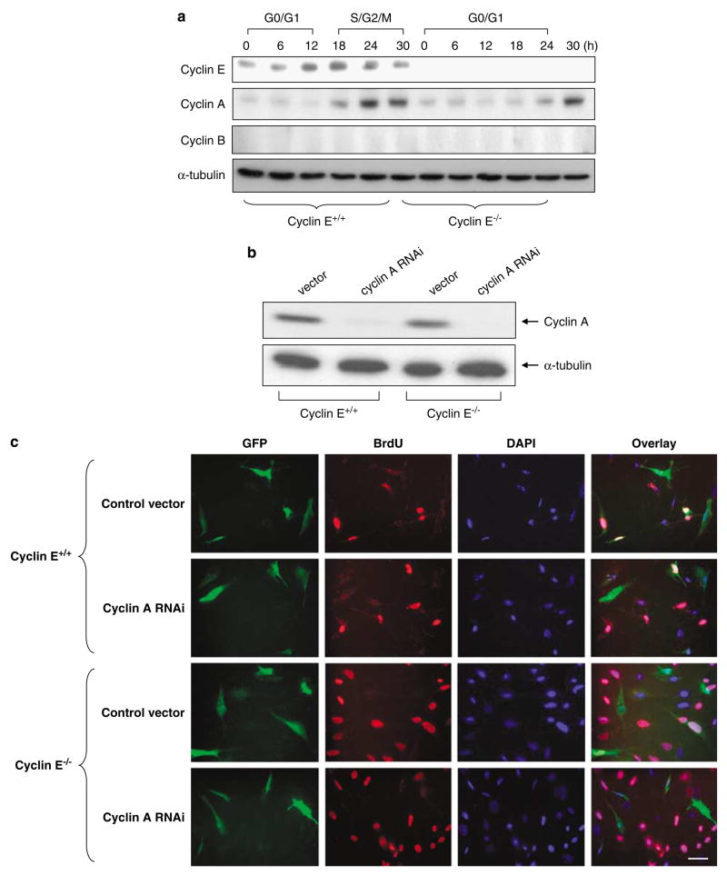
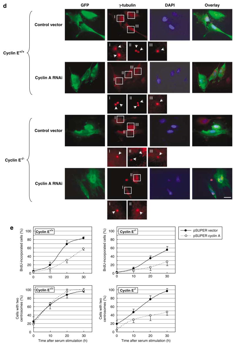
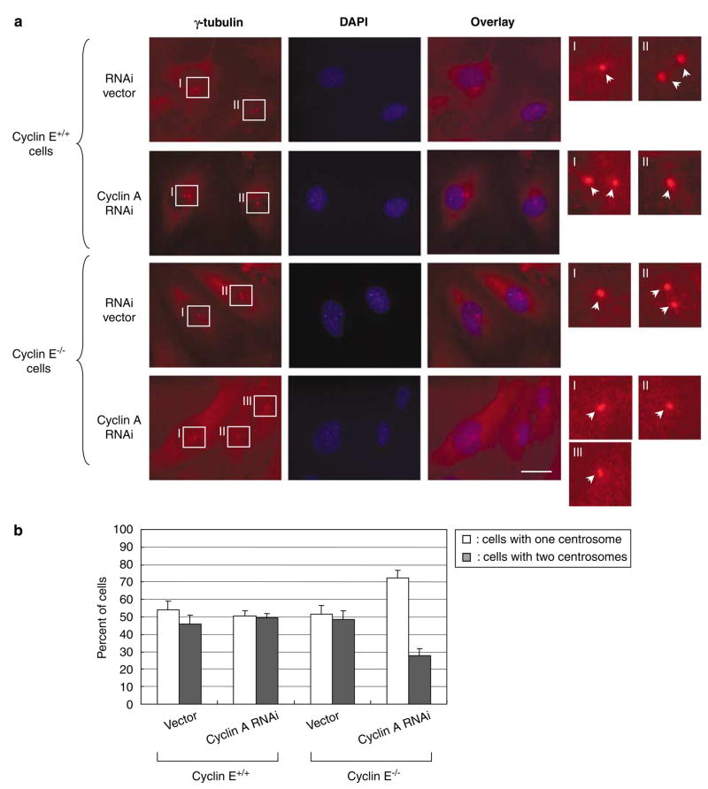
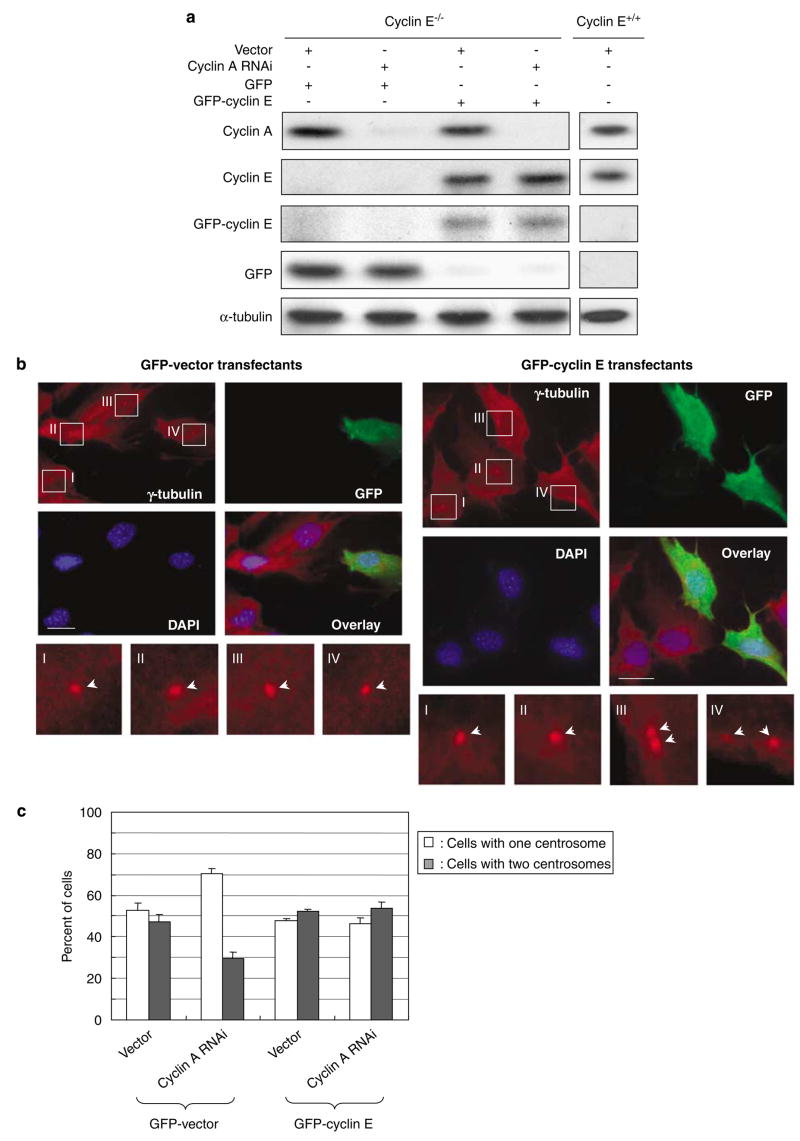
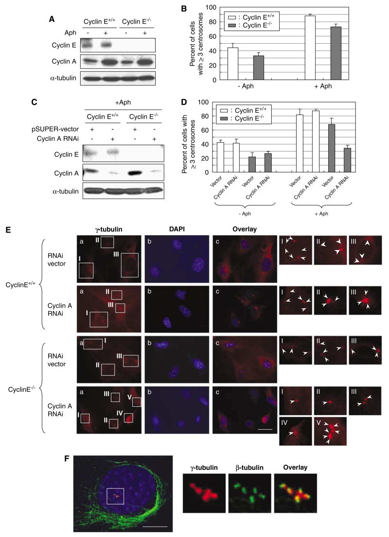
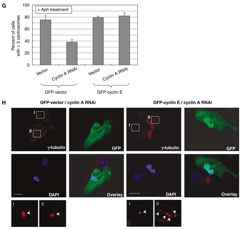
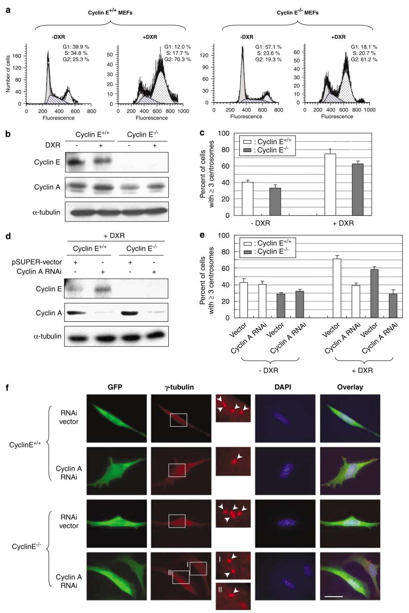
Abnormal amplification of centrosomes, which occurs frequently in cancers, leads to high frequencies of mitotic defect and chromosome segregation error, profoundly affecting the rate of tumor progression. Centrosome amplification results primarily from overduplication of centrosomes, and p53 is involved in the regulation of centrosome duplication partly through controlling the activity of cyclin-dependent kinase (CDK) 2-cyclin E, a kinase complex critical for the initiation of centrosome duplication. Thus, loss or mutational inactivation of p53 leads to an increased frequency of centrosome amplification. Moreover, the status of cyclin E greatly influences the frequency of centrosome amplification in cells lacking functional p53. Here, we dissected the roles of CDK2-associating cyclins, namely cyclins E and A, in centrosome amplification in the p53-negative cells. We found that loss of cyclin E was readily compensated by cyclin A for triggering the initiation of centrosome duplication, and thus the centrosome duplication kinetics was not significantly altered in cyclin E-deficient cells. It has been shown that cells lacking functional p53, when arrested in either early S-phase or late G(2) phase, continue to reduplicate centrosomes, resulting in centrosome amplification. In cells arrested in early S phase, cyclin E, but not cyclin A, is important in centrosome amplification, whereas in the absence of cyclin E, cyclin A is important for centrosome amplification. In late G(2)-arrested cells, cyclin A is important in centrosome amplification irrespective of the cyclin E status. These findings advance our understandings of the mechanisms underlying the numeral abnormality of centrosomes and consequential genomic instability associated with loss of p53 function and aberrant expression of cyclins E and A in cancer cells.


Figures

Figure 1
Figure 1
Analysis of the DNA and centrosome duplication kinetics in cyclins E+/+ and E−/− mouse embryonic fibroblasts (MEFs) expressing DNp53. Cyclins E+/+ and E−/− MEFs expressing DNp53 were serum-starved for 48 h, and then serum-stimulated with the medium containing 20% fetal bovine serum (FBS) and bromodeoxyuridine (BrdU). At indicated time points, cells were fixed and immunostained for incorporated BrdU and γ-tubulin. Representative images of immunofluorescence assay for BrdU incorporation are shown in (a). Scale bar: 40 μm. The rates of BrdU incorporation and centrosome duplication in cyclins E+/+ and E−/− cells were plotted in the graph as average±standard error from three independent experiments (b). For the analysis of centrosomes, >200 γ-tubulin immunostained cells were examined for each time point. In this analysis, cells with amplified (⩾3) centrosomes were excluded from the analysis; it is technically difficult to differentiate unduplicated or duplicated centrosomes in the cells with amplified centrosomes. Determining the rate of centrosome duplication by examining only the cells with one or two centrosomes in the synchronized cells is possible, because the newly duplicated centrosomes do not reduplicate until acquiring the duplication competency. The time course period after serum stimulation in this experiment, there is not sufficient time for the duplicated centrosomes to reestablish the duplication competency. The percentages of the number of cells with two centrosomes among the total number of cells with either one or two centrosomes are plotted in the graph (b) as average±standard error from three independent experiments. Similar results were obtained with the procedure to immunostain centrioles (see Materials and methods section) (data not shown). (c) Determination of doubling times of cyclins E+/+ and E−/− MEFs. Cyclins E+/+ and E−/− cells were plated in the cell culture wells, and the number of cells in the well was counted at every 24 h for 7 days, and plotted in the graph as average±standard error from three independent experiments. (d) Cyclins E+/+ and E−/− MEFs under an optimal growth condition were incubated with BrdU for 1 h, and the percent of cells that had incorporated BrdU was determined, and shown in the graph as average±standard error from three experiments.
Figure 2
Figure 2
Silencing of cyclin A and its effect on centrosome duplication in cyclins E+/+ and E−/− mouse embryonic fibroblasts (MEFs). (a) Cyclin A expression patterns during the cell-cycle progression in cyclins E+/+ and E−/− MEFs. Cyclins E+/+ and E−/− MEFs expressing DNp53 were serum-starved for 48 h, and then serum-stimulated. At indicated time points for a period of 30 h, the lysates were prepared and immunoblotted for cyclins E, A and B and α-tubulin (loading control). Cells were also analysed for cell-cycle phase distributions by flow cytometry, and the respective cell-cycle phases are indicated, in which the majority (>60%) of cells are in the indicated cell-cycle phases (cf. Figure 1b). (b) Cyclins E+/+ and E−/− MEFs were transfected with either a pSUPER plasmid harboring small-interfering RNA (siRNA) sequence specific for cyclin A (pSUPER/cyclin A) or a pSUPER RNA interference (RNAi) vector containing a randomized sequence (pSUPER-vec), along with a puromycin resistance gene as a selection marker. The cells selected in the growth media containing puromycin for 6 days were examined for cyclin A expression by immunoblot analysis. As a loading control, the lysates were probed with anti-α-tubulin antibody. (ce) Cyclins E+/+ and E−/− MEFs were transfected with either pSUPER/cyclin A or pSUPER-vec harboring the randomized sequences along with a plasmid encoding green fluorescent protein (GFP) as a transfection marker. The successfully transfected cells identified by GFP (CycE+/+/CycA RNAi, CycE−/−/CycA RNAi, CycE+/+/vec and CycE−/−/vec cells) were serum-starved for 48 h, and then serum-stimulated in the media containing bromodeoxyuridine (BrdU). At indicated time points, cells were fixed and immunostained with either anti-BrdU or anti-γ-tubulin antibody. Representative images of the BrdU-incorporation assay are shown in (c). Scale bar: 40 μm. Representative images of the γ-tubulin immunostaining are shown in (d). Scale bar: 20 μm. In the magnified images, each centrosome is pointed by an arrow. The rates of BrdU incorporation and centrosome duplication in GFP-positive CycE+/+/CycA RNAi, CycE−/−/CycA RNAi, CycE+/+/vec and CycE−/−/vec cells were plotted in the graph (e). For the analysis of the γ-tubulin immunostained cells,>200 cells for each time point were examined. The rate of centrosome duplication was determined as described in the legend to Figure 1b. The results are shown as the average±standard error from three experiments.
Figure 2
Figure 2
Silencing of cyclin A and its effect on centrosome duplication in cyclins E+/+ and E−/− mouse embryonic fibroblasts (MEFs). (a) Cyclin A expression patterns during the cell-cycle progression in cyclins E+/+ and E−/− MEFs. Cyclins E+/+ and E−/− MEFs expressing DNp53 were serum-starved for 48 h, and then serum-stimulated. At indicated time points for a period of 30 h, the lysates were prepared and immunoblotted for cyclins E, A and B and α-tubulin (loading control). Cells were also analysed for cell-cycle phase distributions by flow cytometry, and the respective cell-cycle phases are indicated, in which the majority (>60%) of cells are in the indicated cell-cycle phases (cf. Figure 1b). (b) Cyclins E+/+ and E−/− MEFs were transfected with either a pSUPER plasmid harboring small-interfering RNA (siRNA) sequence specific for cyclin A (pSUPER/cyclin A) or a pSUPER RNA interference (RNAi) vector containing a randomized sequence (pSUPER-vec), along with a puromycin resistance gene as a selection marker. The cells selected in the growth media containing puromycin for 6 days were examined for cyclin A expression by immunoblot analysis. As a loading control, the lysates were probed with anti-α-tubulin antibody. (ce) Cyclins E+/+ and E−/− MEFs were transfected with either pSUPER/cyclin A or pSUPER-vec harboring the randomized sequences along with a plasmid encoding green fluorescent protein (GFP) as a transfection marker. The successfully transfected cells identified by GFP (CycE+/+/CycA RNAi, CycE−/−/CycA RNAi, CycE+/+/vec and CycE−/−/vec cells) were serum-starved for 48 h, and then serum-stimulated in the media containing bromodeoxyuridine (BrdU). At indicated time points, cells were fixed and immunostained with either anti-BrdU or anti-γ-tubulin antibody. Representative images of the BrdU-incorporation assay are shown in (c). Scale bar: 40 μm. Representative images of the γ-tubulin immunostaining are shown in (d). Scale bar: 20 μm. In the magnified images, each centrosome is pointed by an arrow. The rates of BrdU incorporation and centrosome duplication in GFP-positive CycE+/+/CycA RNAi, CycE−/−/CycA RNAi, CycE+/+/vec and CycE−/−/vec cells were plotted in the graph (e). For the analysis of the γ-tubulin immunostained cells,>200 cells for each time point were examined. The rate of centrosome duplication was determined as described in the legend to Figure 1b. The results are shown as the average±standard error from three experiments.
Figure 3
Figure 3
Analysis of centrosomes in cyclins E+/+ and E−/− mouse embryonic fibroblasts (MEFs) silenced for cyclin A expression under a continual growth condition. CycE+/+/CycA RNAi and CycE−/−/CycA RNAi cells as well as the control CycE+/+/vec and CycE−/−/vec cells were cultured in the complete media, and immunostained with anti-γ-tubulin antibody. The representative immunostaining images are shown in (a). In the magnified images, each centrosome is indicated by an arrow. Scale bar: 20 μm. The ratio of the number of cells with one and two centrosomes in each cell line was determined, and plotted in the graph (b). For each cell line,>200 cells were examined. In this analysis, cells with ⩾3 centrosomes were excluded. The results are shown as the average±standard error from three experiments.
Figure 4
Figure 4
Reintroduction of cyclin E1 abrogates the effect of cyclin A silencing on centrosome duplication. (a) Cyclin E−/− cells were cotransfected with pSUPER/cyclin A and either a pEGFP-C1 vector or a plasmid encoding green fluorescent protein (GFP)-tagged cyclin E1 along with a plasmid encoding a puromycin resistant gene as a rapid selection marker. The lysates prepared from the transfectants were immunoblotted with anti-cyclins A and E1, anti-GFP and anti-α-tubulin antibodies. In parallel, the lysates prepared from cyclin E+/+ mouse embryonic fibroblasts (MEFs) were immunoblotted as a reference for the endogenous level of cyclin E1. (b, c) The transfectants described above were subjected to coimmunostaining with anti-γ-tubulin and anti-GFP antibodies. The representative images of GFP-vector- and GFP-cyclin E1-transfected cells are shown in (b). In the magnified images, each centrosome is indicated by an arrow. Scale bar: 20 μm. The centrosome profiles of the GFP-positive cells were determined, and shown in the graph as the average±standard error from three experiments (c). In this analysis, cells with ⩾3 centrosomes were excluded. For each experiment,>200 cells were examined.
Figure 5
Figure 5
Roles of cyclins A and E in centrosome reduplication in cells arrested by exposure to aphidicolin. (A) Cyclins E+/+ and E−/− mouse embryonic fibroblasts (MEFs) were exposed to Aph (1 μg/ml) for 72 h. The lysates prepared from Aph-treated cells and untreated control cells were immunoblotted with anti-cyclins E and A, and anti-α-tubulin (loading control) antibodies. (B) The Aphtreated and untreated control cells were immunostained for γ-tubulin, and the number of cells with amplified centrosomes were scored. The results are shown in the graph as the average±standard error from three experiments. For each experiment,>200 cells were examined. (C) CycE+/+/CycA RNAi and CycE−/−/CycA RNAi cells as well as the control CycE+/+/vec and CycE−/−/vec cells were treated with Aph for 72 h, and the lysates prepared from Aph-treated cells and untreated control cells were immunoblotted with anticyclins E and A and anti-α-tubulin (loading control) antibodies. (D) The Aph-treated and untreated CycE+/+/CycA RNAi, CycE−/−/CycA RNAi, CycE+/+/vec and CycE−/−/vec cells were immunostained for γ-tubulin, and the number of cells with amplified centrosomes were determined. For each experiment,>200 cells were examined. The results are shown as the average±standard error from three experiments. Representative γ-tubulin immunostaining images are show in (E). In the magnified images, each centrosome is indicated by an arrow. Scale bar: 20 μm. (F)We also examined the centrosomes in the Aph-treated cells by coimmunostaining of γ- and β-tubulins after cold-treatment prior to fixation (see Materials and methods section). By this procedure, centrioles within the centrosome can be visualized. The representative immunostaining images of CycE+/+/CycA RNAi cells after Aph treatment are shown. Scale bar: 5 μm. As described previously, centrosome amplification occurring in cells arrested by Aph is mostly due to centrosome reduplication, and not due to centrosome fragmentation (Tarapore et al., 2001). In addition, by this method, the results for the frequency of centrosome amplification were similar to those by γ-tubulin immunostaining. (G, H) CycE−/−/CycA RNAi and CycE−/−/vec cells were transfected with either a pEGFP-C1 vector or a plasmid encoding green fluorescent protein (GFP)-tagged cyclin E1 (see Figure 4a). The transfectants were then exposed to Aph for 72 h. The Aph-treated and untreated control cells were immunostained for γ-tubulin, and the number of cells with amplified centrosomes were scored. The results are shown in the graph as the average±standard error from three experiments (G). For each experiment, >200 cells were examined. The representative immunostaining images are shown in (H). Scale bar: 20 μm.
Figure 5
Figure 5
Roles of cyclins A and E in centrosome reduplication in cells arrested by exposure to aphidicolin. (A) Cyclins E+/+ and E−/− mouse embryonic fibroblasts (MEFs) were exposed to Aph (1 μg/ml) for 72 h. The lysates prepared from Aph-treated cells and untreated control cells were immunoblotted with anti-cyclins E and A, and anti-α-tubulin (loading control) antibodies. (B) The Aphtreated and untreated control cells were immunostained for γ-tubulin, and the number of cells with amplified centrosomes were scored. The results are shown in the graph as the average±standard error from three experiments. For each experiment,>200 cells were examined. (C) CycE+/+/CycA RNAi and CycE−/−/CycA RNAi cells as well as the control CycE+/+/vec and CycE−/−/vec cells were treated with Aph for 72 h, and the lysates prepared from Aph-treated cells and untreated control cells were immunoblotted with anticyclins E and A and anti-α-tubulin (loading control) antibodies. (D) The Aph-treated and untreated CycE+/+/CycA RNAi, CycE−/−/CycA RNAi, CycE+/+/vec and CycE−/−/vec cells were immunostained for γ-tubulin, and the number of cells with amplified centrosomes were determined. For each experiment,>200 cells were examined. The results are shown as the average±standard error from three experiments. Representative γ-tubulin immunostaining images are show in (E). In the magnified images, each centrosome is indicated by an arrow. Scale bar: 20 μm. (F)We also examined the centrosomes in the Aph-treated cells by coimmunostaining of γ- and β-tubulins after cold-treatment prior to fixation (see Materials and methods section). By this procedure, centrioles within the centrosome can be visualized. The representative immunostaining images of CycE+/+/CycA RNAi cells after Aph treatment are shown. Scale bar: 5 μm. As described previously, centrosome amplification occurring in cells arrested by Aph is mostly due to centrosome reduplication, and not due to centrosome fragmentation (Tarapore et al., 2001). In addition, by this method, the results for the frequency of centrosome amplification were similar to those by γ-tubulin immunostaining. (G, H) CycE−/−/CycA RNAi and CycE−/−/vec cells were transfected with either a pEGFP-C1 vector or a plasmid encoding green fluorescent protein (GFP)-tagged cyclin E1 (see Figure 4a). The transfectants were then exposed to Aph for 72 h. The Aph-treated and untreated control cells were immunostained for γ-tubulin, and the number of cells with amplified centrosomes were scored. The results are shown in the graph as the average±standard error from three experiments (G). For each experiment, >200 cells were examined. The representative immunostaining images are shown in (H). Scale bar: 20 μm.
Figure 6
Figure 6
Roles of cyclins A and E in centrosome reduplication in cells arrested by exposure to doxorubicin (DXR). (a) Cyclins E+/+ and E−/− mouse embryonic fibroblasts (MEFs) were exposed to DXR (0.05 μg/ml) for 48 h, and cell-cycle phase distributions of the cells were analysed by flow cytometry. (b) The lysates prepared from DXR-treated and untreated control cells were immunoblotted with anti-cyclins E and A, and anti-α-tubulin (loading control) antibodies. (c) The DXR-treated and untreated control cells were immunostained for γ-tubulin, and the number of cells with amplified centrosomes were scored. The results are shown in the graph as the average±standard error from three experiments. For each experiment,>200 cells were examined. (d) CycE+/+/CycA RNAi and CycE−/−/CycA RNAi cells as well as the control CycE+/+/vec and CycE−/−/vec cells were treated with DXR for 48 h, and the lysates prepared from the Aph-treated cells and control untreated cells were immunoblotted with anti-cyclins E and, A and anti-α-tubulin (loading control) antibodies. (e) The DXR-treated and untreated control CycE+/+/CycA RNAi, CycE−/−/CycA RNAi, CycE+/+/vec and CycE−/−/vec cells were immunostained for γ-tubulin, and the number of cells with amplified centrosomes were determined. For each experiment,>200 cells were examined. The results are shown in the graph as the average±standard error from three experiments. Representative γ-tubulin immunostaining images are shown in (f). In the magnified images, each centrosome is indicated by an arrow. Scale bar: 20 μm.

References

    1. Balczon R, Bao L, Zimmer WE, Brown K, Zinkowski RP, Brinkley BR. Dissociation of centrosome replication events from cycles of DNA synthesis and mitotic division in hydroxyurea-arrested Chinese hamster ovary cells. J Cell Biol. 1995;130:105–115. - PMC - PubMed
    1. Bennett RA, Izumi H, Fukasawa K. Induction of centrosome amplification and chromosome instability in p53-null cells by transient exposure to subtoxic levels of S-phase-targeting anticancer drugs. Oncogene. 2004;23:6823–6829. - PubMed
    1. Brummelkamp TR, Bernards R, Agami R. A system for stable expression of short interfering RNAs in mammalian cells. Science. 2002;296:550–553. - PubMed
    1. Carroll PE, Okuda M, Horn HF, Biddinger P, Stambrook PJ, Gleich LL, et al. Centrosome hyperamplification in human cancer: chromosome instability induced by p53 mutation and/or Mdm2 overexpression. Oncogene. 1999;18:1935–1944. - PubMed
    1. Coverley D, Laman H, Laskey RA. Distinct roles for cyclins E and A during DNA replication complex assembly and activation. Nat Cell Biol. 2002;4:523–528. - PubMed

Publication types

MeSH terms