. 2006:55:189-97.
doi: 10.3114/sim.55.1.189.

Species of Cercospora associated with grey leaf spot of maize

Affiliations

Species of Cercospora associated with grey leaf spot of maize

Pedro W Crous et al. Stud Mycol. 2006.

Abstract

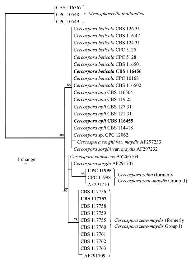
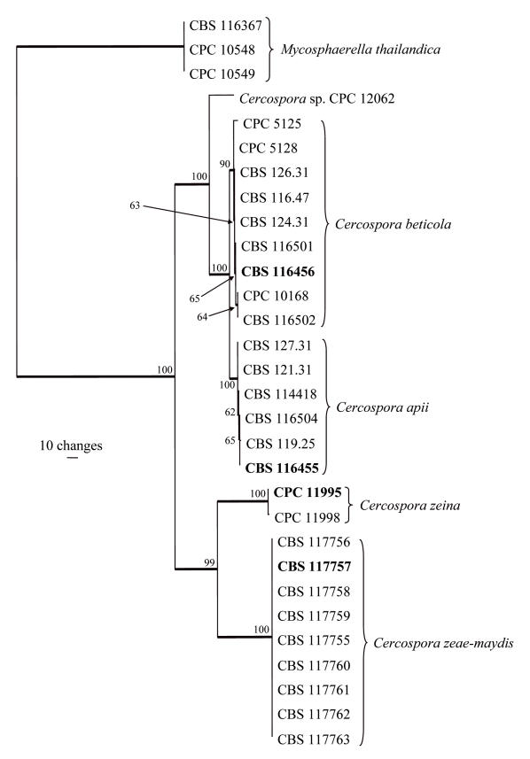
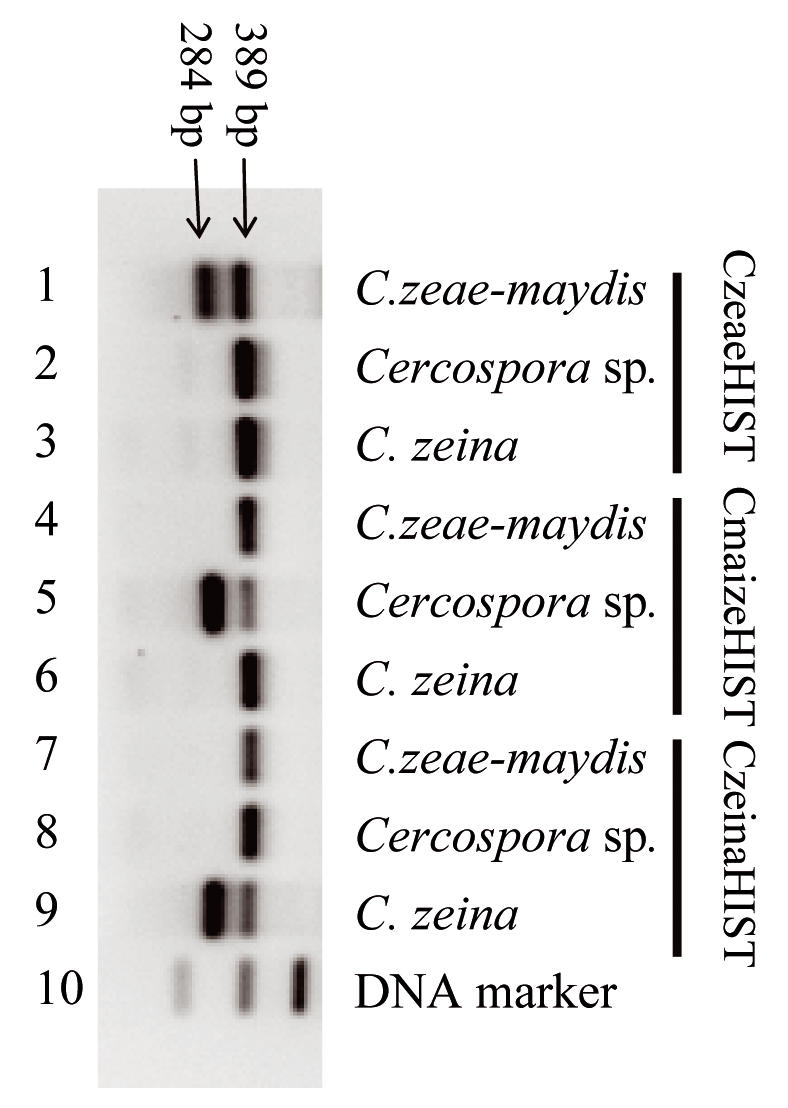
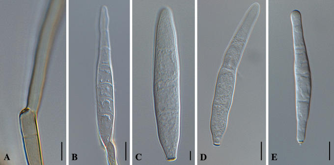
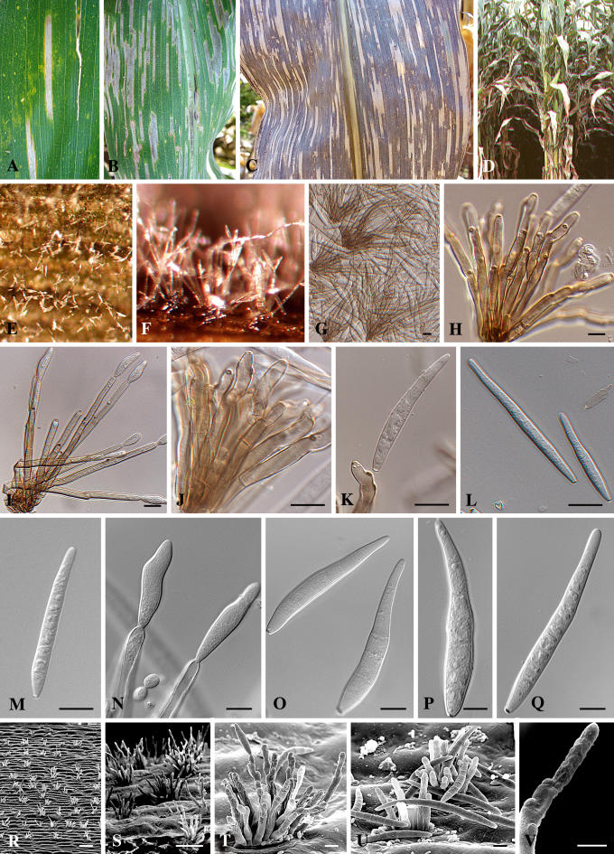
Grey leaf spot is a serious yield-reducing disease of maize (Zea mays) in many parts of the world where this crop is cultivated. The causal organism associated with the disease is Cercospora zeae-maydis. Two potential sibling species have been recognized as Groups I and II. The DNA sequences for the internal transcribed spacers (ITS1 & ITS2), the 5.8S rRNA gene, elongation factor 1-alpha, histone H3, actin and calmodulin gene regions suggest that Groups I and II are two distinct species. Furthermore, Cercospora zeae-maydis (Group I) can be distinguished from C. zeina sp. nov. (Group II) by its faster growth rate on artificial media, the ability to produce cercosporin, longer conidiophores, and broadly fusiform conidia. A PCR-based test that distinguishes the two species was developed using species-specific primers designed from the histone H3 gene.


Figures

Fig. 1.
Fig. 1.
One of six most parsimonious trees obtained from a heuristic search with 100 random taxon additions of the ITS sequence alignment. The scale bar shows a single change, and bootstrap support values from 1000 replicates are shown at the nodes. Thickened lines indicate the strict consensus branches, and ex-type strains are shown in bold print. The tree was rooted to three Mycosphaerella thailandica strains.
Fig. 2.
Fig. 2.
One of two most parsimonious trees obtained from a heuristic search with 100 random taxon additions of the combined ITS, elongation factor 1-alpha, actin, calmodulin and histone H3 sequence alignment. The scale bar shows 10 changes, and bootstrap support values from 1000 replicates are shown at the nodes. Thickened lines indicate the strict consensus branches, and type strains are shown in bold print. The tree was rooted to three Mycosphaerella thailandica strains.
Fig. 3.
Fig. 3.
Identification of C. zeae-maydis, an unidentified Cercospora sp. and C. zeina using the species-specific primers. Lane 10 contains the DNA marker. The 389 bp fragment, which acts as the positive control, is present in all PCR amplifications (lanes 1–9). The species-specific fragment (284 bp) is observed when the amplification reaction contains C. zeae-maydis DNA and primer CzeaeHIST (lane 1, strain CBS 117757), Cercospora sp. DNA and primer CmaizeHIST (lane 5, strain CPC 12062) or C. zeina DNA and primer CzeinaHIST (lane 9, strain CPC 11995).
Fig. 4.
Fig. 4.
Cercospora zeae-maydis. A. Conidiophore with darkened, refractive conidiogenous locus. B. Germinating conidium. C–E. Conidia in vitro. Bars = 10 μm.
Fig. 5.
Fig. 5.
Cercospora zeina. A–C. Close-up of grey leaf spot lesions on maize. D. Heavily infected plant. E–G. Conidiophores fascicles on leaf surface. H–J. Conidiophores. K, N. Conidiogenous cells giving rise to conidia. L–M, O–Q. Conidia. R–U. Scanning electron micrographs of conidiophores and conidia. V. Conidiogenous cell showing thickened loci. Scale bars: G–Q = 10 μm, R = 100 μm, S = 50 μm, T–U = 8 μm, V = 5 μm.

References

    1. Carbone I, Kohn LM (1999). A method for designing primer sets for speciation studies in filamentous ascomycetes. Mycologia 91: 553–556.
    1. Chupp, C. (1954). A monograph of the fungus genus Cercospora. Ithaca, New York. Published by the author.
    1. Corlett M (1991). An annotated list of the published names in Mycosphaerella and Sphaerella. Mycologia Memoir 18, 1–328.
    1. Crous PW (1998). Mycosphaerella spp. and their anamorphs associated with leaf spot diseases of Eucalyptus. Mycologia Memoir 21: 1–170.
    1. Crous PW, Braun U (2003). Mycosphaerella and its anamorphs. 1. Names published in Cercospora and Passalora. CBS Biodiversity Series 1: 1–571.

LinkOut - more resources