. 2007:58:33-56.
doi: 10.3114/sim.2007.58.02.

Delimiting Cladosporium from morphologically similar genera

Affiliations

Delimiting Cladosporium from morphologically similar genera

P W Crous et al. Stud Mycol. 2007.

Abstract

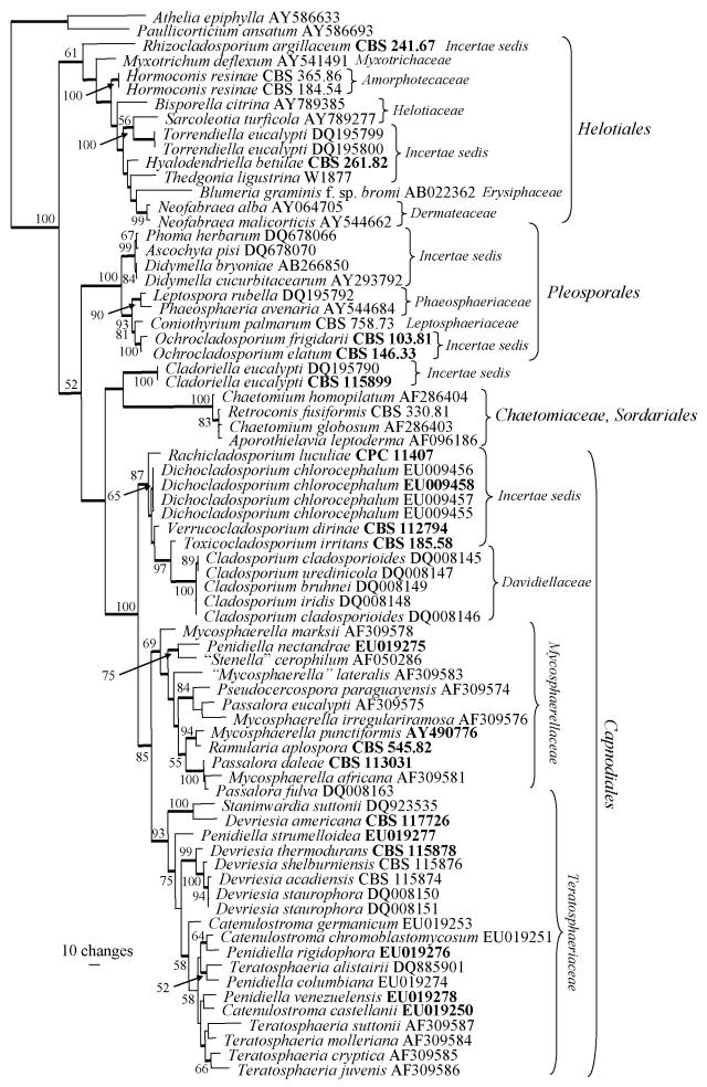
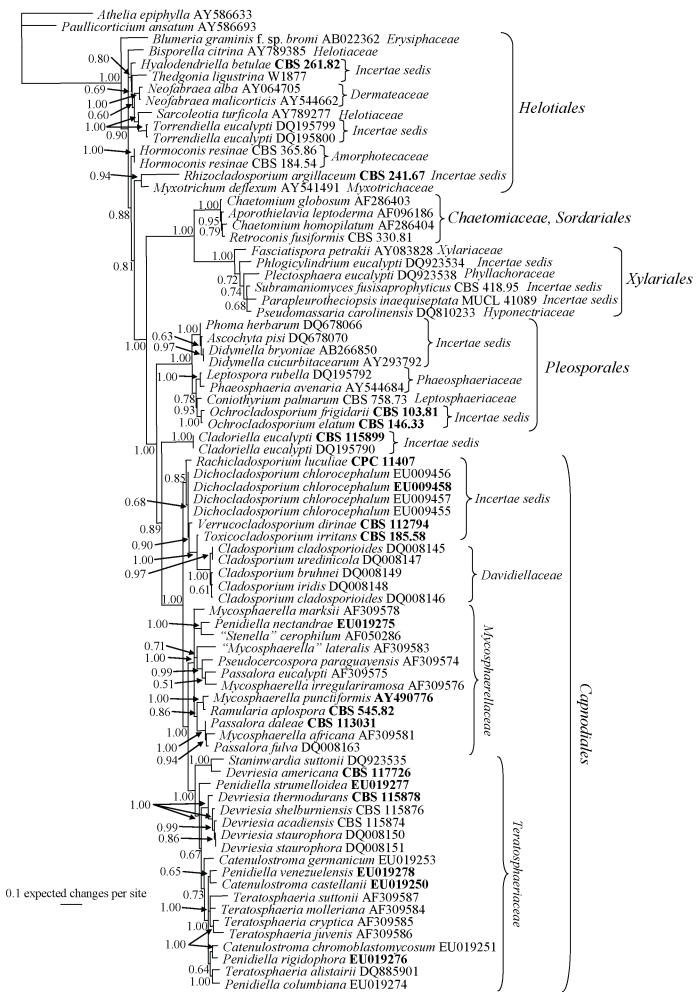
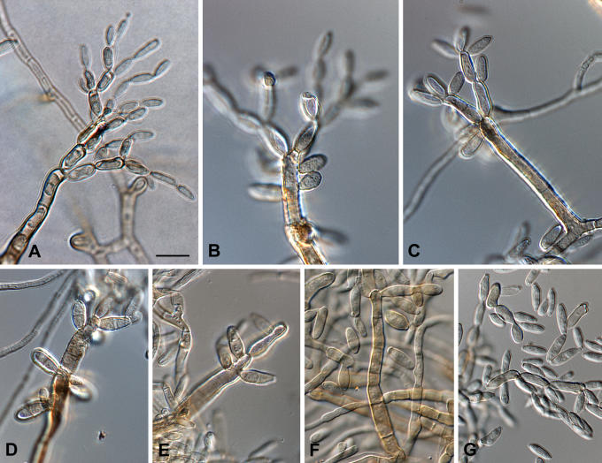
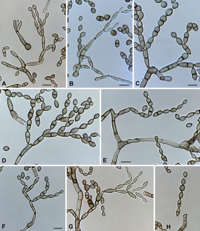
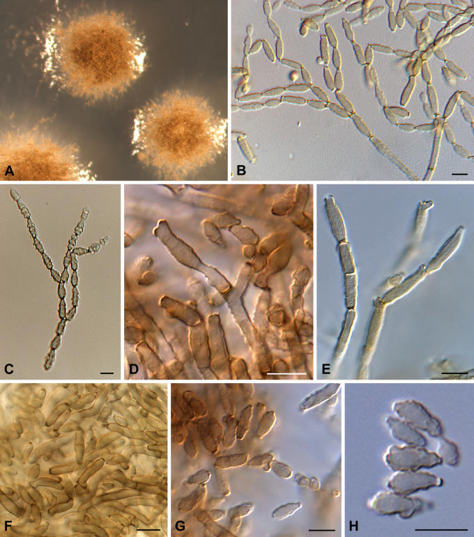
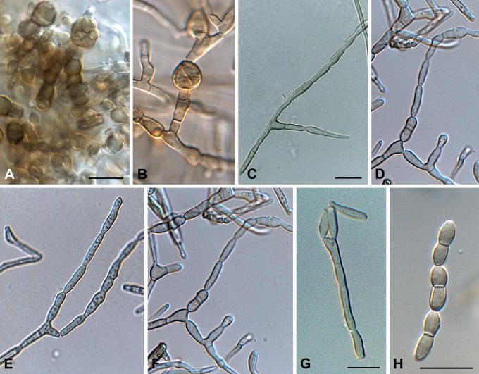
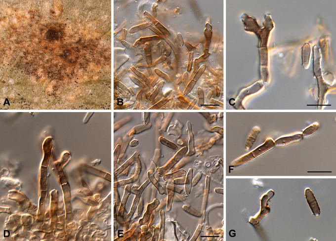
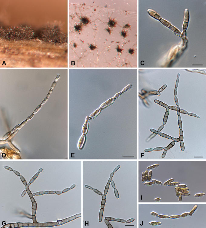
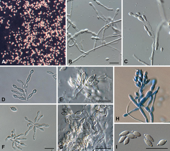
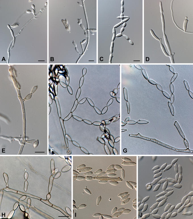
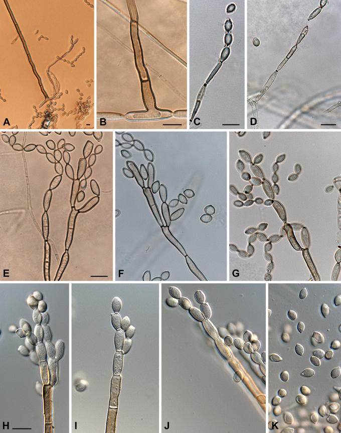
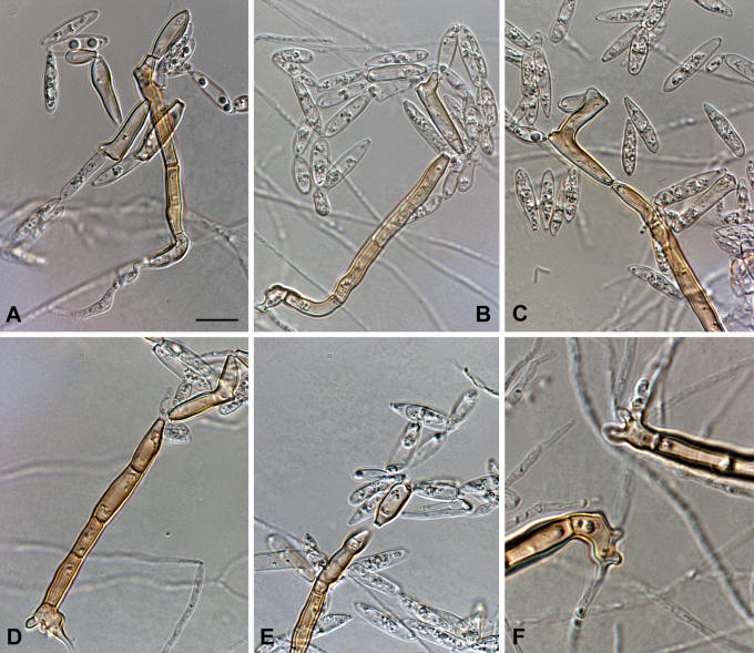
The genus Cladosporium is restricted to dematiaceous hyphomycetes with a coronate scar type, and Davidiella teleomorphs. In the present study numerous cladosporium-like taxa are treated, and allocated to different genera based on their morphology and DNA phylogeny derived from the LSU nrRNA gene. Several species are introduced in new genera such as Hyalodendriella, Ochrocladosporium, Rachicladosporium, Rhizocladosporium, Toxicocladosporium and Verrucocladosporium. A further new taxon is described in Devriesia (Teratosphaeriaceae). Furthermore, Cladosporium castellanii, the etiological agent of tinea nigra in humans, is confirmed as synonym of Stenella araguata, while the type species of Stenella is shown to be linked to the Teratosphaeriaceae (Capnodiales), and not the Mycosphaerellaceae as formerly presumed.


Figures

Fig. 1.
Fig. 1.
One of 44 equally most parsimonious trees obtained from a heuristic search with 100 random taxon additions of the LSU sequence alignment using PAUP v. 4.0b10. The scale bar shows 10 changes, and bootstrap support values from 1 000 replicates are shown at the nodes. Thickened lines indicate the strict consensus branches and ex-type sequences are printed in bold face. The tree was rooted to two sequences obtained from GenBank (Athelia epiphylla AY586633 and Paullicorticium ansatum AY586693).
Fig. 2.
Fig. 2.
Consensus phylogram (50 % majority rule) of 800 trees resulting from a Bayesian analysis of the LSU sequence alignment using MrBayes v. 3.1.2. Bayesian posterior probabilities are indicated at the nodes. Ex-type sequences are printed in bold face. The tree was rooted to two sequences obtained from GenBank (Athelia epiphylla AY586633 and Paullicorticium ansatum AY586693).
Fig. 3.
Fig. 3.
Rachicladosporium luculiae (type material). A-F. Conidiophores with conidial chains, and conidiogenous loci aggregated in the upper region. G. Conidia. Scale bar = 10 μm.
Fig. 4.
Fig. 4.
Toxicocladosporium irritans (type material). A-B, F. Microconidiophores. C-E. Macroconidiophores. G-H. Ramoconidia and conidia. Scale bars = 10 μm.
Fig. 5.
Fig. 5.
Verrucocladosporium dirinae (type material). A. Colonies on MEA. B-C. Conidial chains. D-H. Ramoconidia and conidia. Scale bars = 10 μm.
Fig. 6.
Fig. 6.
Devriesia americana (type material). A-B. Chlamydospore-like structures formed in culture. C-F. Conidiophores giving rise to conidial chains. G-H. Conidia. Scale bars = 10 μm
Fig. 7.
Fig. 7.
Stenella araguata (syntype material, IMI 34905). A. Leaf spot. B. Conidiophore, conidia and verruculose hypha on leaf surface. C-D. Conidiophore with terminal conidiogenous cells. E-G. Ramoconidia and conidia. Scale bars = 10 μm.
Fig. 8.
Fig. 8.
Stenella araguata (CBS 105.75). A-B. Conidiophore fascicles on a pine needle and tap-water agar, respectively. C-D, G. Conidiophores giving rise to conidial chains. E-F, H-J. Conidial chains with ramoconidia and conidia. Scale bars = 10 μm.
Fig. 9.
Fig. 9.
Hyalodendriella betulae (type material). A. Conidiophores on PDA. B-C. Microconidiophores. D-H. Macroconidiophores with fascicles of conidiogenous cells. I. Conidia with darkened, thickened hila. Scale bars = 10 μm.
Fig. 10.
Fig. 10.
Ochrocladosporium elatum (CBS 146.33). A-C, E. Microconidiophores. D. Macro- and microconidiophore. F-H. Macroconidiophores. I-J. Ramoconidia and conidia. Scale bars = 10 μm.
Fig. 11.
Fig. 11.
Ochrocladosporium frigidarii (type material). A. Macro- and microconidiophores. B. Foot cell of macroconidiophore. C-D. Microconidiophore. E-J. Macroconidiophores. K. Conidia. Scale bars = 10 μm.
Fig. 12.
Fig. 12.
Rhizocladosporium argillaceum (type material). A-E. Conidiophores with pigmented ramoconidia and hyaline conidia. F. Rhizoids forming at the foot cells of macroconidiophores. Scale bar = 10 μm.

References

    1. Aptroot A (2006). Mycosphaerella and its anamorphs: 2. Conspectus of Mycosphaerella. CBS Biodiversity Series 5: 1-231.
    1. Arzanlou M, Groenewald JZ, Gams W, Braun U, Shin H-D, Crous PW (2007). Phylogenetic and morphotaxonomic revision of Ramichloridium and allied genera. Studies in Mycology 58: 57-93. - PMC - PubMed
    1. Arx JA von (1974). The Genera of Fungi Sporulating in Pure Culture. 2nd edn. J. Cramer, Vaduz.
    1. Arx JA von (1981). The Genera of Fungi Sporulating in Pure Culture. 3rd edn. J. Cramer, Vaduz.
    1. Arx JA von (1983). Mycosphaerella and its anamorphs. Proceedings of the Koninklijke Nederlandse Akademie van Wetenschappen, Ser. C, 86(1): 15-54.

LinkOut - more resources