. 2007:58:57-93.
doi: 10.3114/sim.2007.58.03.

Phylogenetic and morphotaxonomic revision of Ramichloridium and allied genera

Affiliations

Phylogenetic and morphotaxonomic revision of Ramichloridium and allied genera

M Arzanlou et al. Stud Mycol. 2007.

Abstract

The phylogeny of the genera Periconiella, Ramichloridium, Rhinocladiella and Veronaea was explored by means of partial sequences of the 28S (LSU) rRNA gene and the ITS region (ITS1, 5.8S rDNA and ITS2). Based on the LSU sequence data, ramichloridium-like species segregate into eight distinct clusters. These include the Capnodiales (Mycosphaerellaceae and Teratosphaeriaceae), the Chaetothyriales (Herpotrichiellaceae), the Pleosporales, and five ascomycete clades with uncertain affinities. The type species of Ramichloridium, R. apiculatum, together with R. musae, R. biverticillatum, R. cerophilum, R. verrucosum, R. pini, and three new species isolated from Strelitzia, Musa and forest soil, respectively, reside in the Capnodiales clade. The human-pathogenic species R. mackenziei and R. basitonum, together with R. fasciculatum and R. anceps, cluster with Rhinocladiella (type species: Rh. atrovirens, Herpotrichiellaceae, Chaetothyriales), and are allocated to this genus. Veronaea botryosa, the type species of the genus Veronaea, also resides in the Chaetothyriales clade, whereas Veronaea simplex clusters as a sister taxon to the Venturiaceae (Pleosporales), and is placed in a new genus, Veronaeopsis. Ramichloridium obovoideum clusters with Carpoligna pleurothecii (anamorph: Pleurothecium sp., Chaetosphaeriales), and a new combination is proposed in Pleurothecium. Other ramichloridium-like clades include R. subulatum and R. epichloës (incertae sedis, Sordariomycetes), for which a new genus, Radulidium is erected. Ramichloridium schulzeri and its varieties are placed in a new genus, Myrmecridium (incertae sedis, Sordariomycetes). The genus Pseudovirgaria (incertae sedis) is introduced to accommodate ramichloridium-like isolates occurring on various species of rust fungi. A veronaea-like isolate from Bertia moriformis with phylogenetic affinity to the Annulatascaceae (Sordariomycetidae) is placed in a new genus, Rhodoveronaea. Besides Ramichloridium, Periconiella is also polyphyletic. Thysanorea is introduced to accommodate Periconiella papuana (Herpotrichiellaceae), which is unrelated to the type species, P. velutina (Mycosphaerellaceae).


Figures
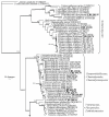
Fig. 1.
Fig. 1.
One of 5 000 equally most parsimonious trees obtained from a heuristic search with simple taxon additions of the LSU sequence alignment using PAUP v. 4.0b10. The scale bar shows 10 changes; bootstrap support values from 1 000 replicates are shown at the nodes. Thickened lines indicate the strict consensus branches and ex-type sequences are printed in bold face. The tree was rooted to two sequences obtained from GenBank (Athelia epiphylla AY586633 and Paullicorticium ansatum AY586693).
Fig. 1.
Fig. 1.
One of 5 000 equally most parsimonious trees obtained from a heuristic search with simple taxon additions of the LSU sequence alignment using PAUP v. 4.0b10. The scale bar shows 10 changes; bootstrap support values from 1 000 replicates are shown at the nodes. Thickened lines indicate the strict consensus branches and ex-type sequences are printed in bold face. The tree was rooted to two sequences obtained from GenBank (Athelia epiphylla AY586633 and Paullicorticium ansatum AY586693).
Fig. 2.
Fig. 2.
Consensus phylogram (50 % majority rule) of 1 500 trees resulting from a Bayesian analysis of the LSU sequence alignment using MrBayes v. 3.1.2. Bayesian posterior probabilities are indicated at the nodes. Ex-type sequences are printed in bold face. The tree was rooted to two sequences obtained from GenBank (Athelia epiphylla AY586633 and Paullicorticium ansatum AY586693).
Fig. 2.
Fig. 2.
Consensus phylogram (50 % majority rule) of 1 500 trees resulting from a Bayesian analysis of the LSU sequence alignment using MrBayes v. 3.1.2. Bayesian posterior probabilities are indicated at the nodes. Ex-type sequences are printed in bold face. The tree was rooted to two sequences obtained from GenBank (Athelia epiphylla AY586633 and Paullicorticium ansatum AY586693).
Fig. 3.
Fig. 3.
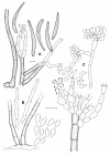
Periconiella velutina (CBS 101948). A-B. Macronematous conidiophores with short branches in the upper part. C. Sympodially proliferating conidiogenous cell with darkened and slightly thickened scars. D. Conidia. Scale bar = 10 μm.
Fig. 4.
Fig. 4.
Periconiella arcuata (CBS 113477). A-B. Sympodially proliferating conidiogenous cells with darkened, thickened and cone-shaped scars. C-E. Macronematous conidiophores with loose branches in the upper part. F-I. Conidia. Scale bar = 10 μm.
Fig. 5.
Fig. 5.
Periconiella levispora (CBS 873.73). A-C. Conidial apparatus at different stages of development, which gives rise to macronematous conidiophores with dense branches in the upper part. D. Sympodially proliferating conidiogenous cells with darkened and somewhat protruding scars. E-F. Conidia with truncate base and darkened hilum. Scale bar = 10 μm.
Fig. 6.
Fig. 6.
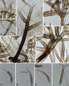
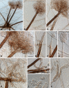
A. Pseudovirgaria hyperparasitica (CBS 121739 = CPC 10753). B. Periconiella levispora (CBS 873.73). Scale bar = 10 μm.
Fig. 7.
Fig. 7.
A. Periconiella arcuata (CBS 113477). B. Myrmecridium schulzeri (CBS 325.74). C. Thysanorea papuana (CBS 212.96). Scale bars = 10 μm.
Fig. 8.
Fig. 8.
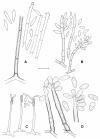
Ramichloridium apiculatum (CBS 156.59). A-C. Macronematous conidiophores with sympodially proliferating conidiogenous cells, which give rise to a conidium-bearing rachis with crowded and prominent scars. D. Conidia. Scale bar = 10 μm.
Fig. 9.
Fig. 9.
Ramichloridium australiense (CBS 121710). A-C. Macronematous conidiophores with thick-walled and warted subtending hyphae. D. Sympodially proliferating conidiogenous cell, which results in a short rachis with darkened and slightly thickened scars. E. Conidia. Scale bar = 10 μm.
Fig. 10.
Fig. 10.
A. Ramichloridium australiense (CBS 121710). B. Ramichloridium brasilianum (CBS 283.92). C. Radulidium subulatum (CBS 405.76). D. Rhodoveronaea varioseptata (CBS 431.88). Scale bar = 10 μm.
Fig. 11.
Fig. 11.
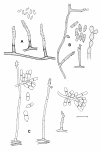
Ramichloridium musae (CBS 365.36). A. Conidiophores with loose branches. B-D. Sympodially proliferating conidiogenous cells, resulting in a long conidium-bearing rachis. E. Rachis with hardly prominent, slightly darkened scars. F. Conidia. Scale bars = 10 μm.
Fig. 12.
Fig. 12.
Ramichloridium biverticillatum (CBS 335.36). A-B. Profusely branched and biverticillate conidiophores. C. Sympodially proliferating conidiogenous cells, which give rise to a conidium-bearing rachis with crowded, slightly pigmented and thickened scars. D. Conidia. Scale bar = 10 μm.
Fig. 13.
Fig. 13.
Ramichloridium brasilianum (CBS 283.92). A-B. Macronematous conidiophores with sympodially proliferating conidiogenous cells, resulting in a conidium-bearing rachis. C. Rachis with crowded and slightly pigmented scars. D. Conidia. Scale bar = 10 μm.
Fig. 14.
Fig. 14.
Ramichloridium cerophilum (CBS 103.59). A-C. Conidial apparatus at different stages of development, resulting in macronematous conidiophores and sympodially proliferating conidiogenous cells. D-E. Formation of secondary conidia. F. Conidia. Scale bar = 10 μm.
Fig. 15.
Fig. 15.
Ramichloridium indicum (CBS 171.96). A-B. Macronematous conidiophores. C-E. Sympodially proliferating conidiogenous cells, resulting in a conidium-bearing rachis with pigmented and thickened scars. F. Conidia. Scale bar = 10 μm.
Fig. 16.
Fig. 16.
Ramichloridium strelitziae (CBS 121711). A-C. Conidial apparatus at different stages of development, resulting in macronematous conidiophores and sympodially proliferating conidiogenous cells. D-E. Rachis with crowded, slightly pigmented, thickened, circular scars. F. Conidia. Scale bars = 10 μm.
Fig. 17.
Fig. 17.
A. Ramichloridium strelitziae (CBS 121711). B. Veronaea japonica (CBS 776.83). C. Veronaeopsis simplex (CBS 588.66). Scale bar = 10 μm.
Fig. 18.
Fig. 18.
Zasmidium cellare (CBS 146.36). A-D. Micronematous conidiophores with terminal, integrated conidiogenous cells. E. Conidiogenous cell with pigmented, thickened and refractive scars. F-G. Primary and secondary conidia. Scale bar = 10 μm.
Fig. 19.
Fig. 19.
Rhinocladiella anceps (CBS 181.65). A. Macronematous conidiophores. B-D. Conidial apparatus at different stages of development, resulting in semi-micronematous conidiophores and sympodially proliferating conidiogenous cells. E. Conidiogenous loci. F. Conidia. Scale bars = 10 μm.
Fig. 20.
Fig. 20.
Rhinocladiella basitona (CBS 101460). A-B. Semi-micronematous conidiophores with verticillate branching pattern. C-D. Sympodially proliferating conidiogenous cells, giving rise to a long rachis with slightly prominent, truncate conidium-bearing denticles. E. Intercalary conidiogenous cell. F. Conidia. Scale bars = 10 μm.
Fig. 21.
Fig. 21.
Rhinocladiella fasciculata (CBS 132.86). A. Conidiophores. B. Sympodially proliferating conidiogenous cells, which give rise to a long rachis with slightly prominent, unthickened scars. C. Conidia. D-E. Synanamorph consisting of conidiogenous cells with percurrent proliferation. F. Conidia. Scale bars = 10 μm.
Fig. 22.
Fig. 22.
Rhinocladiella mackenziei (CBS 368.92). A. Intercalary conidiogenous cell. B-E. Semi-micronematous conidiophores and sympodially proliferating conidiogenous cells, resulting in a rachis with slightly prominent, unthickened scars. F. Conidia. Scale bar = 10 μm.
Fig. 23.
Fig. 23.
Thysanorea papuana (CBS 212.96). A. Intercalary conidiogenous cell. B-I. Semi-micronematous conidiophores and sympodially proliferating conidiogenous cells, resulting in a rachis with prominent conidium bearing denticles. J-K. Microcyclic conidiation observed in slide cultures. L. Conidia. Scale bar = 10 μm.
Fig. 24.
Fig. 24.
Thysanorea papuana (CBS 212.96), periconiella-like synanamorph. A. Macronematous conidiophores. B-C. Conidiophores with dense apical branches. D. Branches with different levels of branchlets. E-I. Conidiogenous cells at different stages of development; sympodially proliferating conidiogenous cells give rise to a denticulate rachis. J-K. Conidia. Scale bars = 10 μm.
Fig. 25.
Fig. 25.
Veronaea botryosa (CBS 254.57). A-C. Semi-micronematous conidiophores and sympodially proliferating conidiogenous cells. D-E. Rachis with crowded and flat scars. F-G. Microcyclic conidiation. H. Conidia. Scale bars = 10 μm.
Fig. 26.
Fig. 26.
Veronaea compacta (CBS 268.75). A-B. Semi-micronematous conidiophores and sympodially proliferating conidiogenous cells. C-D. Rachis with hardly prominent denticles. E. Conidia. Scale bar = 10 μm.
Fig. 27.
Fig. 27.
Veronaea japonica (CBS 776.83). A. Intercalary conidiogenous cells. B-D. Semi-micronematous conidiophores and sympodially proliferating conidiogenous cells. E. Conidia. F. Thick-walled, dark brown hyphal cells. Scale bar = 10 μm.
Fig. 28.
Fig. 28.
Pleurothecium obovoideum (CBS 209.95). A-C. Conidial apparatus consisting of conidiophores with sympodially proliferating conidiogenous cells as seen in slide cultures of ca. 14 d. D. Short chain of conidia. E-G. Sympodially proliferating conidiogenous cells, resulting in a short rachis with subcylindrical to cylindrical denticles. H. Conidia. Scale bar = 10 μm.
Fig. 29.
Fig. 29.
Myrmecridium schulzeri (CBS 325.74). A. Macronematous conidiophores. B. Inflated basal cells visible in some conidiophores. C-E. Conidial apparatus at different stages of development, resulting in macronematous conidiophores and sympodially proliferating conidiogenous cells. F-G. Rachis with scattered, pimple-shaped denticles. H. Conidia. Scale bars: A =100 μm, B-H = 10 μm.
Fig. 30.
Fig. 30.
Myrmecridium flexuosum (CBS 398.76). A-C. Conidial apparatus at different stages of development, resulting in macronematous conidiophores with sympodially proliferating conidiogenous cells. D-H. Sympodially proliferating conidiogenous cells giving rise to a flexuose conidium-bearing rachis with pimple-shaped denticles. I. Conidia. Scale bar = 10 μm.
Fig. 31.
Fig. 31.
Pseudovirgaria hyperparasitica (CBS 121739). A-D. Conidial apparatus at different stages of development; conidiogenous cells with geniculate proliferation. E. Conidia. Scale bar = 10 μm.
Fig. 32.
Fig. 32.
Radulidium subulatum (CBS 405.76). A-B. Macronematous conidiophores with sympodially proliferating conidiogenous cells, resulting in a conidium-bearing rachis. C-D. Rachis with crowded, blunt conidium-bearing denticles. E. Conidia. Scale bar = 10 μm.
Fig. 33.
Fig. 33.
Radulidium epichloës (CBS 361.63). A-C. Conidial apparatus at different stages of development, resulting in semi-micronematous conidiophores and sympodially proliferating conidiogenous cells. D. Rachis with crowded, blunt conidium-bearing denticles. E-F. Conidiophores with acute branches in the lower part. G. Conidia. Scale bar = 10 μm.
Fig. 34.
Fig. 34.
Rhodoveronaea varioseptata (CBS 431.88). A-D. Macronematous conidiophores with sympodially proliferating conidiogenous cells, resulting in conidium bearing rachis with slightly prominent conidium-bearing denticles. E-F. Conidia with minute marginal frill. Scale bar = 10 μm.
Fig. 35.
Fig. 35.
Veronaeopsis simplex (CBS 588.66). A-C. Conidial apparatus at different stages of development, resulting in semi-micronematous conidiophores and sympodially proliferating conidiogenous cells. D-E. Rachis with crowded, prominent denticles. F. Intercalary conidiogenous cells. G. Conidia. Scale bar = 10 μm.

References

    1. Al-Hedaithy SSA, Jamjoom ZAB, Saeed ES (1988). Cerebral phaeohyphomycosis caused by Fonsecaea pedrosoi in Saudi-Arabia. Acta Pathologica Microbiologica Scandinaviae 96Suppl. 3: 94-100. - PubMed
    1. Aptroot A (2006). Mycosphaerella and its anamorphs: 2. Conspectus of Mycosphaerella. CBS Biodiversity Series 5: 1-231.
    1. Braun U (1998). A monograph of Cercosporella, Ramularia and allied genera (phytopathogenic hypomycetes). Vol. 2. IHW-Verlag, Eching.
    1. Braun U, Crous PW, Dugan F, Groenewald JZ, Hoog GS de (2003). Phylogeny and taxonomy of cladosporium-like hyphomycetes, including Davidiella gen. nov., the teleomorph of Cladosporium s. str. Mycological Progress 2: 3-18.
    1. Campbell CK, Al-Hedaithy SSA (1993). Phaeohyphomycosis of the brain caused by Ramichloridium mackenziei sp. nov. in middle eastern countries. Journal of Medical and Veterinary Mycology 31: 325-332.

LinkOut - more resources