. 2007:58:219-34.
doi: 10.3114/sim.2007.58.08.

Molecular analysis and pathogenicity of the Cladophialophora carrionii complex, with the description of a novel species

Affiliations

Molecular analysis and pathogenicity of the Cladophialophora carrionii complex, with the description of a novel species

G S de Hoog et al. Stud Mycol. 2007.

Abstract

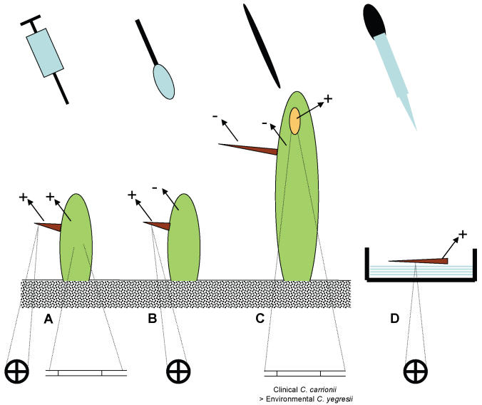
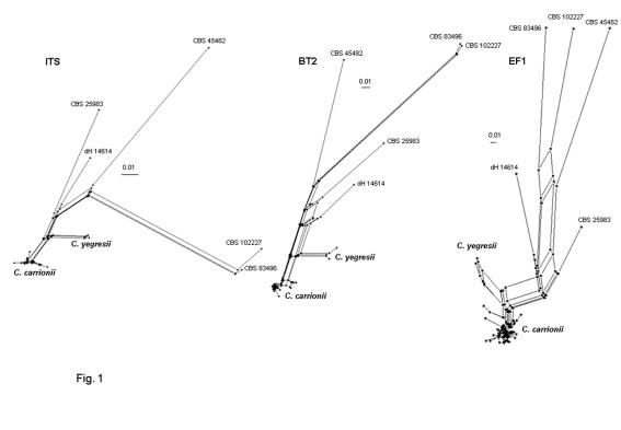
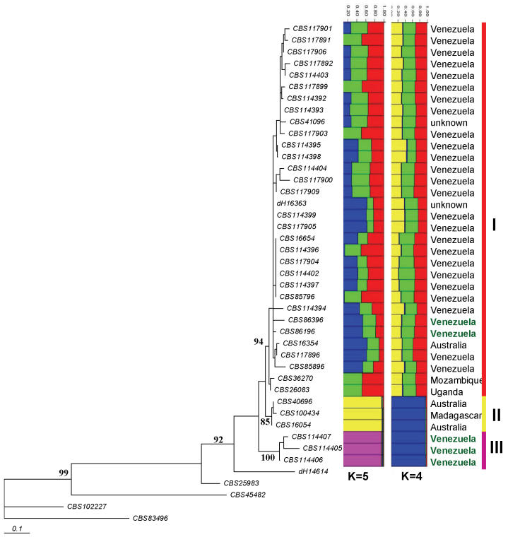
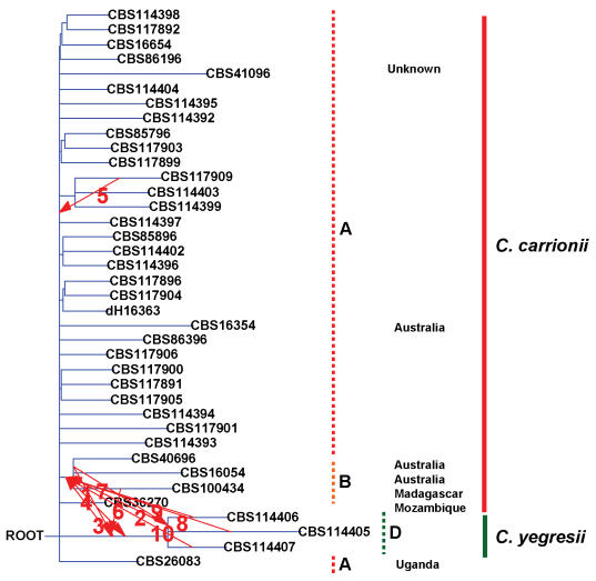
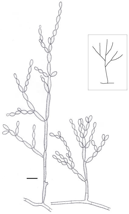
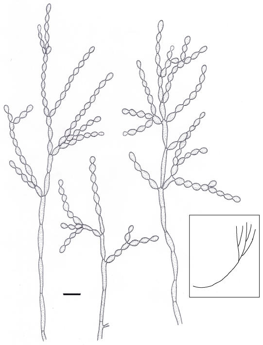
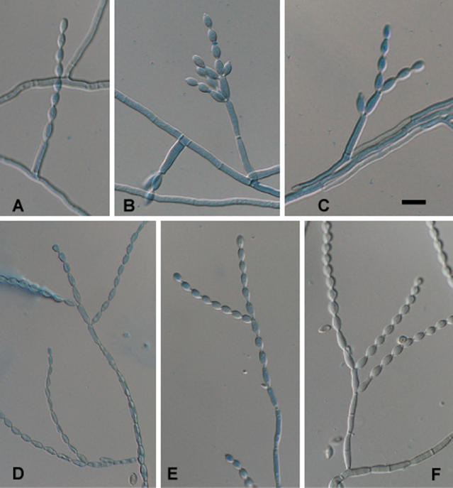
Cladophialophora carrionii is one of the four major etiologic agents of human chromoblastomycosis in semi-arid climates. This species was studied using sequence data of the internal transcribed spacer region of rDNA, the partial beta-tubulin gene and an intron in the translation elongation factor 1-alpha gene, in addition to morphology. With all genes a clear bipartition was observed, which corresponded with minute differences in conidiophore morphology. A new species, C. yegresii, was introduced, which appeared to be, in contrast to C. carrionii, associated with living cactus plants. All strains from humans, and a few isolates from dead cactus debris, belonged to C. carrionii, for which a lectotype was designated. Artificial inoculation of cactus plants grown from seeds in the greenhouse showed that both fungi are able to persist in cactus tissue. When reaching the spines they produce cells that morphologically resemble the muriform cells known as the "invasive form" in chromoblastomycosis. The tested clinical strain of C. carrionii proved to be more virulent in cactus than the environmental strain of C. yegresii that originated from the same species of cactus, Stenocereus griseus. The muriform cell expressed in cactus spines can be regarded as the extremotolerant survival phase, and is likely to play an essential role in the natural life cycle of these organisms.


Figures

Fig. 1.
Fig. 1.
Diagram of inoculation experiments with results. A. Inoculation of young cactus; B. Superficial application of young cactus; C. Traumatic application of mature cactus, with brown resulting scar; D. Superficial application of mature spines. Indications +/- refer to positive resp. negative results of re-isolated strains. Lower line: circles represent production of muriform cells, filaments represent hyphal growth.
Fig. 2.
Fig. 2.
Split decomposition of the C. carrionii complex using SplitsTree with uncorrected (P-value) distances. Nodes are shown only with different genotypes; hence EF1 shows the largest number of nodes. Extensive reticulation is noted in all loci.
Fig. 3.
Fig. 3.
Phylogenetic tree (Neighbour-joining) of the C. carrionii complex based on EF1 (with grouping I-III) using the same strains of Fig. 1, generated using the HKY+G model. The model was calculated using ML in MrAic software. Bootstrap cut-off = 80 %. CBS 834.96 was taken as outgroup. Columns were generated with Structure software hypothesising K = 4 and K = 5, and alleles independent. Geographical origins in black refer to isolates from humans (chromoblastomycosis); origins in green refer to isolates from plant material.
Fig. 4.
Fig. 4.
Reticulogram of South American strains of Cladophialophora species and strains from other continents (mentioned) constructed with T-rex software. ITS rDNA (with grouping A, B, D) was used as species tree and compared with the ß-tubulin gene tree. First 10 reticulations are shown with numbers.
Fig. 5.
Fig. 5.
Microscopic morphology of C. carrionii, strain CBS 160.54. Conidiophore erect, i.e. mostly arising at 90° from creeping hypha (sketch). Scale bar = 10 μm.
Fig. 6.
Fig. 6.
Microscopic morphology of C. yegresii, strain CBS 114405. Conidiophore ascending, i.e. mostly emerging from hyphal end that is gradually growing upwards to become a conidiophore (sketch). Scale bar = 10 μm.
Fig. 7.
Fig. 7.
Conidial morphology in selected branches of (upper row: A-C) C. carrionii, strain CBS 260.83; (lower row: D-F) C. yegresii, strain CBS 114405. In this respect the two species are identical. Scale bar = 10 μm.

References

    1. Abliz P, Fukushima K, Takizawa K, Nishimura K (2004). Specific oligonucleotide primers for identification of Cladophialophora carrionii, a causative agent of chromoblastomycosis. Journal of Clinical Microbiology 42: 404-407. - PMC - PubMed
    1. Ahmed AOA, Adelmann D, Fahal A, Verbrugh H, Belkum A van, Hoog GS de (2002). Environmental occurrence of Madurella mycetomatis, the major agent of human eumycetoma in Sudan. Journal of Clinical Microbiology 40: 1031-1036. - PMC - PubMed
    1. Ahrens J, Graybill JR, Abishawl A, Tio FO, Rinaldi MG (1989). Experimental murine chromomycosis mimicking chronic progressive human disease. American Journal of Tropical Medicine and Hygiene 40: 651-658. - PubMed
    1. Albornoz MB, Marin C de, Iwatsu T (1982). Estudio epidemiologico de un area endemica para cromomicosis en el Estado Falcon. Investigación Clínica 23: 219-228.
    1. Bandelt HJ, Dress AW (1992). Split decomposition: a new and useful approach to phylogenetic analysis of distance data. Molecular Phylogenetics and Evolution 1: 242-252. - PubMed

LinkOut - more resources