A mathematical model platform for optimizing a multiprojection breast imaging system
- PMID: 18491528
- PMCID: PMC2673621
- DOI: 10.1118/1.2885367
A mathematical model platform for optimizing a multiprojection breast imaging system
Abstract
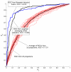
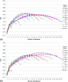
Multiprojection imaging is a technique in which a plurality of digital radiographic images of the same patient are acquired within a short interval of time from slightly different angles. Information from each image is combined to determine the final diagnosis. Projection data are either reconstructed into slices as in the case of tomosynthesis or analyzed directly as in the case of multiprojection correlation imaging technique, thereby avoiding reconstruction artifacts. In this study, the authors investigated the optimum geometry of acquisitions of a multiprojection breast correlation imaging system in terms of the number of projections and their total angular span that yield maximum performance in a task that models clinical decision. Twenty-five angular projections of each breast from 82 human subjects in our breast tomosynthesis database were each supplemented with a simulated 3 mm mass. An approach based on Laguerre-Gauss channelized Hotelling observer was developed to assess the detectability of the mass in terms of receiver operating characteristic (ROC) curves. Two methodologies were developed to integrate results from individual projections into one combined ROC curve as the overall figure of merit. To optimize the acquisition geometry, different components of acquisitions were changed to investigate which one of the many possible configurations maximized the area under the combined ROC curve. Optimization was investigated under two acquisition dose conditions corresponding to a fixed total dose delivered to the patient and a variable dose condition, based on the number of projections used. In either case, the detectability was dependent on the number of projections used, the total angular span of those projections, and the acquisition dose level. In the first case, the detectability approximately followed a bell curve as a function of the number of projections with the maximum between 8 and 16 projections spanning angular arcs of about 23 degrees-45 degrees, respectively. In the second case, the detectability increased with the number of projections approaching an asymptote at 11-17 projections for an angular span of about 45 degrees. These results indicate the inherent information content of the multi-projection image data reflecting the relative role of quantum and anatomical noise in multiprojection breast imaging. The optimization scheme presented here may be applied to any multiprojection imaging modalities and may be extended by including reconstruction in the case of digital breast tomosynthesis and breast computed tomography.
Figures


References
-
- Samei E., Flynn M. J., and Eyler W. R., “Relative influence of quantum and anatomical noise on the detection of subtle lung nodules in chest radiographs,” Radiology 213, 727–734 (1999). - PubMed
-
- Bochud F. O., Verdun F. R., Hessler C., and Valley J. -F., “Detectability of radiological images: The influence of anatomical noise,” Proc. SPIE PSISDG10.1117/12.206845 2436, 156–164 (1995). - DOI
-
- Zhou L., Oldan J., Fisher P., and Gindi G., “Low-contrast lesion detection in tomosynthetic breast imaging using a realistic breast phantom,” Proc. SPIE PSISDG10.1117/12.652343 6142, 61425A (2006). - DOI
Publication types
MeSH terms
Grants and funding
LinkOut - more resources
Full Text Sources
Medical

