

Automated detection of microcalcification clusters for digital breast tomosynthesis using projection data only: a preliminary study
- PMID: 18491543
- PMCID: PMC2811555
- DOI: 10.1118/1.2885366
Automated detection of microcalcification clusters for digital breast tomosynthesis using projection data only: a preliminary study
Abstract
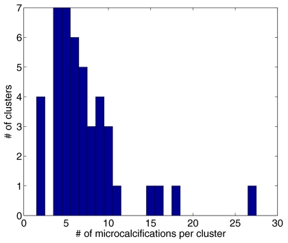
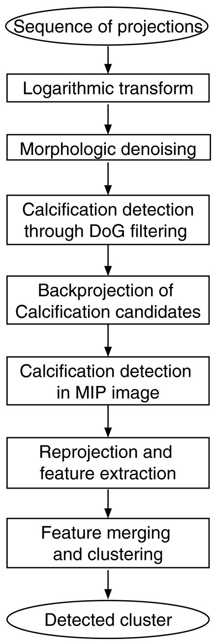
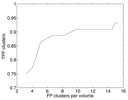
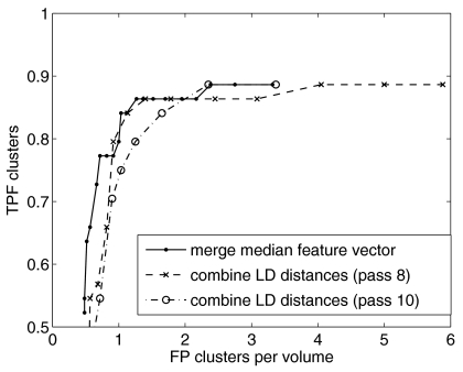
Digital breast tomosynthesis (DBT) is a promising modality for breast imaging in which an anisotropic volume image of the breast is obtained. We present an algorithm for computerized detection of microcalcification clusters (MCCs) for DBT. This algorithm operates on the projection views only. Therefore it does not depend on reconstruction, and is computationally efficient. The algorithm was developed using a database of 30 image sets with microcalcifications, and a control group of 30 image sets without visible findings. The patient data were acquired on the first DBT prototype at Massachusetts General Hospital. Algorithm sensitivity was estimated to be 0.86 at 1.3 false positive clusters, which is below that of current MCC detection algorithms for full-field digital mammography. Because of the small number of patient cases, algorithm parameters were not optimized and one linear classifier was used. An actual limitation of our approach may be that the signal-to-noise ratio in the projection images is too low for microcalcification detection. Furthermore, the database consisted of predominantly small MCC. This may be related to the image quality obtained with this first prototype.
Figures

References
-
- Wu T., Moore R. H., and Kopans D. B., “Voting strategy for artifact reduction in digital breast tomosynthesis,” Med. Phys. MPHYA6 33, 2641–2671 (2006). - PubMed
-
- Rafferty E. A., “Digital mammography: Novel applications,” Radiol. Clin. North Am. RCNAAU 45, 831–834 (2007). - PubMed
-
- Wu T., Stewart A., Stanton M., McCauley T., Phillips W., Kopans D. B., Moore R. H., Eberhard J. W., Opsahl-Ong B., Niklason L., and Williams M. B., “Tomographic mammography using a limited number of low-dose cone-beam projection images,” Med. Phys. MPHYA610.1118/1.1543934 30, 365–380 (2003). - DOI - PubMed
Publication types
MeSH terms
Grants and funding
LinkOut - more resources
Full Text Sources
Other Literature Sources
Medical
