. 2009 Apr;19(2):214-23.
doi: 10.1111/j.1750-3639.2008.00176.x. Epub 2008 May 19.

A lack of amyloid beta plaques despite persistent accumulation of amyloid beta in axons of long-term survivors of traumatic brain injury

Affiliations

A lack of amyloid beta plaques despite persistent accumulation of amyloid beta in axons of long-term survivors of traumatic brain injury

Xiao-Han Chen et al. Brain Pathol. 2009 Apr.

Abstract

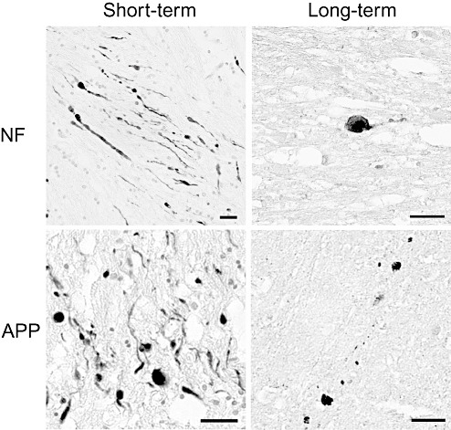
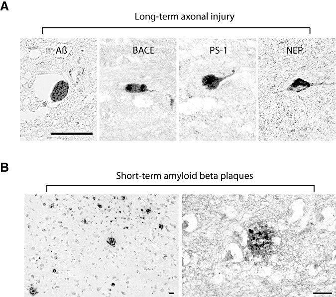
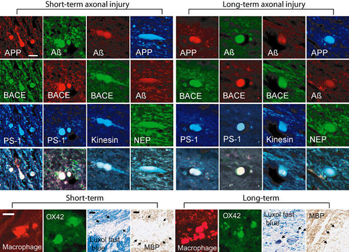
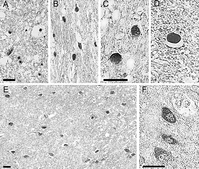
Traumatic brain injury (TBI) is a risk factor for developing Alzheimer's disease (AD). Additionally, TBI induces AD-like amyloid beta (Abeta) plaque pathology within days of injury potentially resulting from massive accumulation of amyloid precursor protein (APP) in damaged axons. Here, progression of Abeta accumulation was examined using brain tissue from 23 cases with post-TBI survival of up to 3 years. Even years after injury, widespread axonal pathology was consistently observed and was accompanied by intra-axonal co-accumulations of APP with its cleavage enzymes, beta-site APP cleaving enzyme and presenilin-1 and their product, Abeta. However, in marked contrast to the plaque pathology noted in short-term cases post TBI, virtually no Abeta plaques were found in long-term survivors. A potential mechanism for Abeta plaque regression was suggested by the post-injury accumulation of an Abeta degrading enzyme, neprilysin. These findings fail to support the premise that progressive plaque pathology after TBI ultimately results in AD.


Figures

Figure 1
Figure 1
Axonal pathology. Representative immunoreactivity for neurofilament (NF) and amyloid precursor protein (APP) demonstrating axonal bulbs and varicosities in both short‐ and long‐term survival cases of traumatic brain injury. Scale bar = 50 µm.
Figure 2
Figure 2
Long‐term intra‐axonal pathology and short‐term amyloid plaques. (A) Axonal bulb immunoreactivity for amyloid β (Aβ) (6F/3D) antibody, β‐site APP cleaving enzyme (BACE), presenilin‐1 (PS‐1) and neprilysin in long‐term survival cases. (B) Aβ plaques found in short‐term survival cases (4G8 antibody). Scale bar = 50 µm.
Figure 3
Figure 3
Axonal bulb accumulations, macrophages/microglia and myelin. Top panels: representative triple labeling of amyloid precursor protein (APP), amyloid β (Aβ), β‐site APP cleaving enzyme (BACE), presenilin‐1 (PS‐1), kinesin, neprilysin (NEP), and APP within both short‐ and long‐term survival cases. Immunoreactivity gives the typical appearance of these proteins accumulating within axonal bulbs in the subcortical white matter. 13335 and Amy33 antibodies demonstrate Aβ within axonal bulbs. Neprilysin can be found co‐localizing with APP and Aβ. Scale bar = 50 µm. Bottom Panels: Immunofluorescence demonstrated macrophages/ microglia in the brain tissue in short‐ and long‐term survival cases detected by antibodies OX42 and macrophage inflammatory protein. Luxol fast blue and immunostaining for myelin basic protein (SMI 94) revealed patchy loss of immunoreactivity in the subcortical white matter in both short‐ and long‐term survival patients. Scale bar = 50 µm.
Figure 4
Figure 4
Neprilysin staining. Photomicrographs demonstrating immunoreactivity to neprilysin representative of positive cases found up to 8 months post injury. (A and B) Clusters of positively stained axons are seen within the subcortical white matter. Note the classic appearance of halos surrounding the swollen axon terminals. This phenomenon occurs secondary to dehydration of the tissue during processing. (C and D) Swollen axon bulbs seen at higher magnification. (E) Clusters of soma within the subcortical white matter demonstrating positive neprilysin staining. (F) Somal neprilysin staining seen at a higher magnification. Note the staining has a granulated quality. All scale bars = 50 µm.

Similar articles

Cited by

References

    1. Adams JH, Doyle D, Ford I, Gennarelli TA, Graham DI, McLellan DR (1989) Diffuse axonal injury in head injury: definition, diagnosis and grading. Histopathology 15:49–59. - PubMed
    1. Adams JH, Graham DI (1976) The relationship between ventricular fluid pressure and the neuropathology of raised intracranial pressure. Neuropathol Appl Neurobiol 2:323–332.
    1. Adams JH, Graham DI, Gennarelli TA, Maxwell WL (1991) Diffuse axonal injury in non‐missile head injury. J Neurol Neurosurg Psychiatry 54:481–483. - PMC - PubMed
    1. Adams JH, Graham DI, Jennett B (2000) The neuropathology of the vegetative state after an acute brain insult. Brain 123:1327–1338. - PubMed
    1. Adams JH, Graham DI, Jennett B (2001) The structural basis of moderate disability after traumatic brain damage. J Neurol Neurosurg Psychiatry 71:521–524. - PMC - PubMed

Publication types