Comparative Study
. 2008 May 20:9:26.
doi: 10.1186/1471-2121-9-26.

Straw blood cell count, growth, inhibition and comparison to apoptotic bodies

Affiliations
Comparative Study

Straw blood cell count, growth, inhibition and comparison to apoptotic bodies

Yonnie Wu et al. BMC Cell Biol. .

Abstract

Background: Mammalian cells transform into individual tubular straw cells naturally in tissues and in response to desiccation related stress in vitro. The transformation event is characterized by a dramatic cellular deformation process which includes: condensation of certain cellular materials into a much smaller tubular structure, synthesis of a tubular wall and growth of filamentous extensions. This study continues the characterization of straw cells in blood, as well as the mechanisms of tubular transformation in response to stress; with specific emphasis placed on investigating whether tubular transformation shares the same signaling pathway as apoptosis.

Results: There are approximately 100 billion, unconventional, tubular straw cells in human blood at any given time. The straw blood cell count (SBC) is 45 million/ml, which accounts for 6.9% of the bloods dry weight. Straw cells originating from the lungs, liver and lymphocytes have varying nodules, hairiness and dimensions. Lipid profiling reveals severe disruption of the plasma membrane in CACO cells during transformation. The growth rates for the elongation of filaments and enlargement of rabbit straw cells is 0.6 approximately 1.1 (microm/hr) and 3.8 (microm(3)/hr), respectively. Studies using apoptosis inhibitors and a tubular transformation inhibitor in CACO2 cells and in mice suggested apoptosis produced apoptotic bodies are mediated differently than tubular transformation produced straw cells. A single dose of 0.01 mg/kg/day of p38 MAPK inhibitor in wild type mice results in a 30% reduction in the SBC. In 9 domestic animals SBC appears to correlate inversely with an animal's average lifespan (R2 = 0.7).

Conclusion: Straw cells are observed residing in the mammalian blood with large quantities. Production of SBC appears to be constant for a given animal and may involve a stress-inducible protein kinase (P38 MAPK). Tubular transformation is a programmed cell survival process that diverges from apoptosis. SBCs may be an important indicator of intrinsic aging-related stress.


Figures
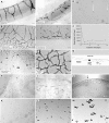
Figure 1
Figure 1
Straw blood cells in air and in solution. Straw cells originating from mouse, human and other animals. A. Straw cells derived from mouse epithelia, lymphocyte, liver and lung cells. B Human filamentous straw cells, C. Human smooth straw cells, D. Pig lung straw cells, E. Straw cells in solution; visible dark dots have diameters near 1 μm (arrows). F. Turnover of straw cells incubated at 37°C in vitro (▼ Human blood, ▲ Rabbit sera). G. Carton presentation of a straw cell and its components. H. Synchronized rabbit straw cells in air. Straw cells losing filamentous extensions were incubated in sterile water at day-0. I. Emergence of new filaments at day-1. J. Emergence of rabbit straw cell network in 5 days (dark arrow), emergence of aggregated cell bodies in solution (white arrow). K. Emergence of rabbit regular cells in 6 days, 12 days (L) and 14 days (M). Space bar equals to 10 μm.
Figure 2
Figure 2
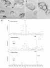
Production of straw cells in vitro. Straw cells from dehydration induced CACO2 cells in vitro. A. Normal CACO2 cells, B. SC filaments in solution and in air, C. Surface and total lipids from normal and induced cells.
Figure 3
Figure 3
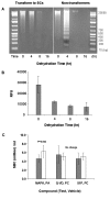
Comparison of tubular transformation with apoptotic bodies.A. No appearance of DNA ladder (left panel) in CACO2 cells during a successful tubular transformation induced by dehydration. DNA ladder did appear (right panel) in CACO2 cells during a failed tubular transformation B. Measurement of caspase-3/7 activity during dehydration induced tubular transformation in CACO2 cells. C. Inhibition of tubular transformation by small molecules in vivo. Blood samples are counted from a single dose sc at 24 hr.
Figure 4
Figure 4
The relationship between SBC and expected animal lifespan. The Figure is plotted using data from Table 1.
Figure 5
Figure 5
Identification of proteins involved in tubular transformation and regeneration.A. Purified tubular proteins on SDS/PAGE in10% acrylamide gel from MCF-7 and CACO2 cells that were transformed into straw cells. B. Regeneration of regular cells from straw cells, three proteins at 63, 57, and 52 KD were abundantly expressed in the early stage of the process.

References

    1. Wu Y, Laughlin RC, Henry DC, Krueger DE, Hudson JS, Kuan CY, He J, Reppert J, Tomkins JP. Naturally occurring and stress induced tubular structures from mammalian cells, a survival mechanism. BMC Cell Biology. 2007;8:36. doi: 10.1186/1471-2121-8-36. - DOI - PMC - PubMed
    1. Hengartner MO, Ellis RE, Horvitz HR. C. elegans cell survival gene ced-9 protects cells from programmed cell death. Nature. 1992;356:494–9. doi: 10.1038/356494a0. - DOI - PubMed
    1. Kerr JFR, Wyllie AH, Currie AR. Apoptosis: A basic biological phenomenon with wide-ranging implications in tissue kinetics. Br J Cancer. 1972;26:239–57. - PMC - PubMed
    1. Leist M, and Jäättelä M. Four deaths and a funeral: From caspases to alternative mechanisms. Nat Rev Mol Cell Biol. 2001;2:589–98. doi: 10.1038/35085008. - DOI - PubMed
    1. Hengartner MO. The biochemistry of apoptosis. Nature. 2000;407:770–6. doi: 10.1038/35037710. - DOI - PubMed

Publication types

MeSH terms

LinkOut - more resources