. 2008 May 20:8:8.
doi: 10.1186/1475-2867-8-8.

Reduced paxillin expression contributes to the antimetastatic effect of 4-hydroxycoumarin on B16-F10 melanoma cells

Affiliations

Reduced paxillin expression contributes to the antimetastatic effect of 4-hydroxycoumarin on B16-F10 melanoma cells

Marco A Velasco-Velázquez et al. Cancer Cell Int. .

Abstract

Background: 4-Hydroxycoumarin (4-HC) is a coumarin that lacks anticoagulant activity. 4-HC affects the cytoskeletal stability and decreases cell adhesion and motility of the melanoma cell line B16-F10. Together with integrins and other cytoskeletal proteins, paxillin participates in the regulation of cell adhesion and motility, acting as an adapter protein at focal adhesions. The present study determined the participation of paxillin in the reported effects of 4-HC and analyzed the role of paxillin in the formation of melanoma metastases.

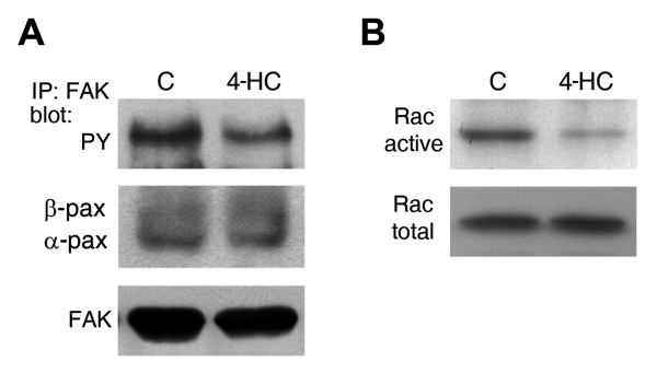
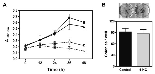
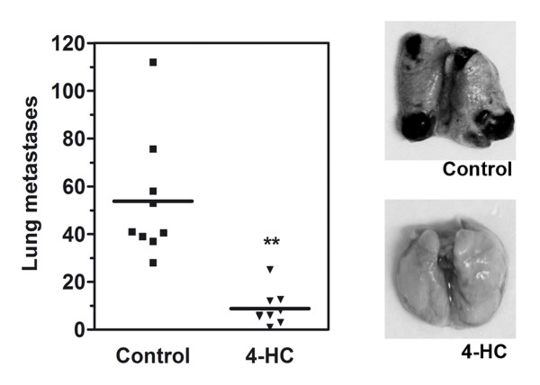
Results: 4-HC decreased protein and mRNA levels of alpha- and beta-paxillin isoforms in B16-F10 cells. Paxillin downregulation correlated with an inadequate translocation of paxillin to focal adhesions and a reduced phosphotyr118-paxillin pool. Consequently, 4-HC altered paxillin-mediated signaling, decreasing the phosphorylation of FAK and the level of GTP-bound Rac-1. These results partially explain the mechanism of the previously reported effects of 4-HC. Additionally, we studied the effect of 4-HC on metastatic potential of B16-F10 cells through experimental metastasis assays. In vitro treatment of cells with 4-HC inhibited their capability to originate pulmonary metastases. 4-HC did not affect cell proliferation or survival, demonstrating that its antimetastatic effect is unrelated to changes on cell viability. We also studied the importance of paxillin in metastasis by transfecting melanoma cells with paxillin-siRNA. Transfection produced a modest reduction on metastatic potential, indicating that: i) paxillin plays a role as inducer of melanoma metastasis; and ii) paxillin downregulation is not sufficient to explain the antimetastatic effect of 4-HC. Therefore, we evaluated other changes in gene expression by differential display RT-PCR analysis. Treatment with 4-HC produced a downregulation of Adhesion Regulating Molecule-1 (ARM-1), which correlated with a decreased adhesion of melanoma cells to lung slides.

Conclusion: This study shows that reduced paxillin expression is associated with the impaired cell adhesion and motility seen in 4-HC-treated cells and partially contributes to the antimetastatic effect of 4-HC. In contrast, the role of ARM-1 reduced expression in the effects of 4-HC is still to be clarified. The antimetastatic effect of 4-HC suggests that this compound, or others with similar mode of action, might be useful for the development of adjuvant therapies for melanoma.


Figures

Figure 1
Figure 1
Paxillin expression, phosphorylation and localization in 4-HC-treated cells. A. Representative immunoblot showing decreased levels of both paxillin isoforms in 4-HC-treated cells. As loading control, β-tubulin was detected in the same membrane. Quantification of paxillin expression (graph) was performed by densitometric analysis from 4 independent experiments. On each experiment, the value of the band corresponding to α-paxillin in control lane was arbitrarily set to 1. Paxillin/tubulin ratios (mean ± SE) were reduced in 4-HC-treated cells (open bars in graph) when compared with control cells (closed bars). B. Semiquantitative RT-PCR using primers for pan-paxillin (upper panel), β-paxillin (middle panel) or GAPDH (lower panel). One representative experiment from 3 is shown. C. Representative photomicrographs from immunofluorescences performed with anti-paxillin antibody. In control cells (left), paxillin was located in focal adhesions around the cell periphery; this pattern was modified by the treatment with 4-HC (right). Scale bar = 20 μm. D. The level of phosphotyr118-paxillin (pY118) was also diminished by 4-HC. We were only able to detect phosphorylation in the α isofom of paxillin. One representative experiment from 3 is shown.
Figure 2
Figure 2
Effect of 4-HC on the basal activation of FAK and Rac-1. A. Analysis of samples immunoprecipitated with anti-FAK antibody showed that basal tyrosine phosphorylation of FAK (upper panel) was decreased by 4-HC. The same membrane was stripped and reprobed with anti-paxillin and anti-FAK antibodies. We found no changes on the FAK-bound paxillin levels (middle panel). One representative experiment from 3 is shown. B. The active form of Rac-1 was purified using pull-down assays and then probed with anti-Rac-1 antibody (upper panel). Detection of Rac-1 from total lysates (lower panel) showed that the difference in the amount of active Rac-1 is not associated to changes on its expression. One representative experiment from 2 is shown.
Figure 3
Figure 3
Effect of 4-HC on cell proliferation and survival. A. B16-F10 cells were treated with 500 μM 4-HC (triangles) or 0.75% ethanol (control cells; squares) and viable cells were quantified every 12 h using the neutral red accumulation assay. Experiments performed in presence of 10% FBS (solid lines) showed that cell proliferation was unaffected by 4-HC (P > 0.05, ANOVA). When cells were treated in serum-free medium (dashed lines), the initial cell number was unaltered neither by time nor by 4-HC. Values are mean ± SE obtained from 3 independent experiments. B. Clonogenic assay were performed by seeding 4-HC-treated cells at a density of 20 cells/cm2. After incubation for 8 d, the generated colonies were stained and counted. There was no difference in the colony number produced by 4-HC-treated cells or control cells. Values in the graph are mean ± SE obtained from 3 independent experiments. The upper picture shows examples of the results.
Figure 4
Figure 4
Effect of 4-HC on the formation of experimental metastases. Melanoma cells were treated in vitro with 500 μM 4-HC for 24 h, and then injected into the tail vein of C57BL/6 mice. Two weeks later, mice were sacrificed and the macroscopic pulmonary tumors were counted. Mice injected with 4-HC-treated cells (triangles) showed a 7-fold decrease in the number of lung tumors compared with the mice that received control B16-F10 cells (squares). ** P = 0.002 (Student's t test with Welch's correction). The pictures on the right show examples of lungs from mice injected with either control or 4-HC-treated cells.
Figure 5
Figure 5
Effect of paxillin-siRNA on the formation of experimental metastases. A. Immunoblot showing that paxillin expression was reduced in B16-F10 cells transfected with paxillin-siRNA (pax). Densitometric quantification (graph) show that the decrements on paxillin/tubulin ratios (mean ± SE, n = 3) in cells transfected with paxillin-siRNA (open bars) were similar to those produced by 4-HC. B. The participation of paxillin in metastasis formation was studied by injecting into mice cells transfected with either paxillin- or control-siRNA. Paxillin downregulation produced a 1.6-fold reduction in the number of experimental pulmonary metastases. * P = 0.04 (Student's t test). The pictures on the right show examples of lungs from mice injected with siRNAs-trated cells.
Figure 6
Figure 6
ARM-1 expression and adhesion to lung sections by cells treated with 4-HC. A. The effect of 4-HC on ARM-1 expression was evaluated by real time-PCR. 4-HC inhibited ARM-1 mRNA expression. Values are represented as ratios of ARM-1/GAPDH ± SD. B. B16-F10 cells were treated as described previously, and then incubated with lung sections during 30 min to allow their adhesion. The preparations were stained and photographed. Photomicrographs (400X) show that control cells (left) were able to adhere to lung sections; in contrast, 4-HC-treated cells (right) displayed impaired adhesion. C. Quantification of adhered cells (mean ± SE) from 5 independent experiments. On each experiment, 10 fields were counted. ***P < 0.0001 (Student's t test).

References

    1. Bogenrieder T, Herlyn M. Axis of evil: molecular mechanisms of cancer metastasis. Oncogene. 2003;22:6524–6536. doi: 10.1038/sj.onc.1206757. - DOI - PubMed
    1. Hood JD, Cheresh DA. Role of integrins in cell invasion and migration. Nat Rev Cancer. 2002;2:91–100. doi: 10.1038/nrc727. - DOI - PubMed
    1. Geiger B, Bershadsky A, Pankov R, Yamada K. Transmembrane extracellular matrix-cytoskeleton crosstalk. Nat Rev Mol Cell Biol. 2001;2:793–805. doi: 10.1038/35099066. - DOI - PubMed
    1. Ridley AJ, Schwartz MA, Burridge K, Firtel RA, Ginsberg MH, Borisy G, Parsons JT, Horwitz AR. Cell migration: integrating signals from front to back. Science. 2003;302:1704–1709. doi: 10.1126/science.1092053. - DOI - PubMed
    1. Hehlgans S, Haase M, Cordes N. Signaling via integrins: implications for cell survival and anticancer strategies. Biochim Biophys Acta. 2007;1775:163–180. - PubMed