Comparative Study
. 2008 May 21:5:19.
doi: 10.1186/1742-2094-5-19.

Glucagon-like peptide 1 receptor stimulation reverses key deficits in distinct rodent models of Parkinson's disease

Affiliations
Comparative Study

Glucagon-like peptide 1 receptor stimulation reverses key deficits in distinct rodent models of Parkinson's disease

Alexander Harkavyi et al. J Neuroinflammation. .

Abstract

Background: It has recently become apparent that neuroinflammation may play a significant role in Parkinson's disease (PD). This is also the case in animal paradigms of the disease. The potential neuroprotective action of the glucagon-like peptide 1 receptor (GLP-1R) agonist exendin-4 (EX-4), which is protective against cytokine mediated apoptosis and may stimulate neurogenesis, was investigated In paradigms of PD.

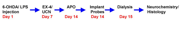
Methods: Two rodent 'models' of PD, 6-hydroxydopamine (6-OHDA) and lipopolysaccaride (LPS), were used to test the effects of EX-4. Rats were then investigated in vivo and ex vivo with a wide range of behavioural, neurochemical and histological tests to measure integrity of the nigrostriatal system.

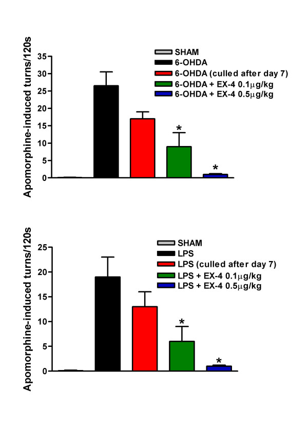
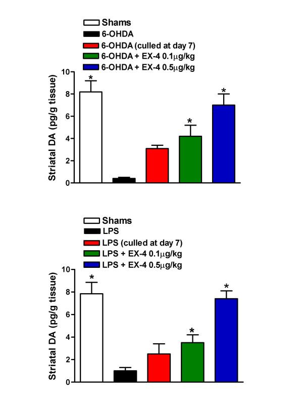
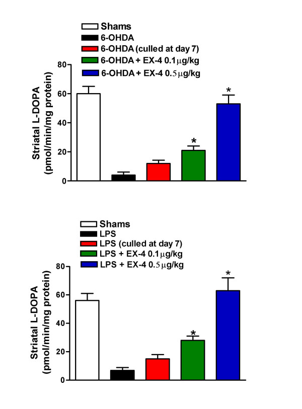
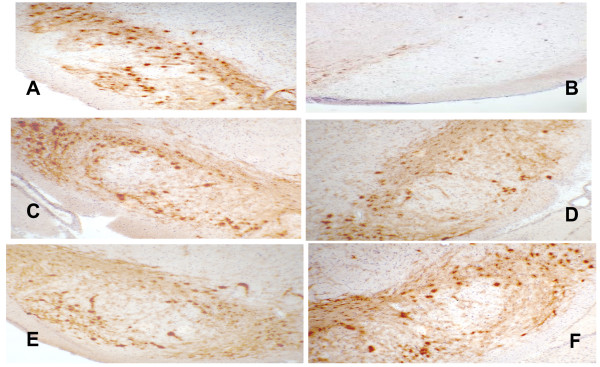
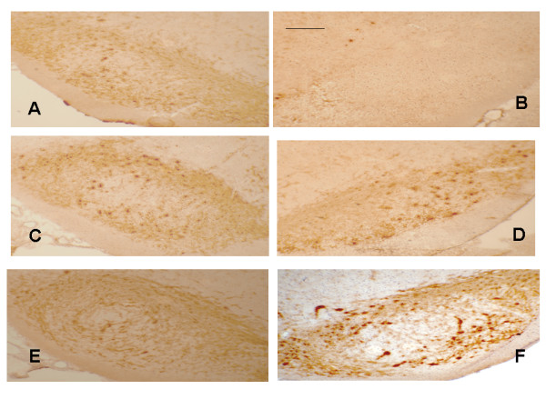
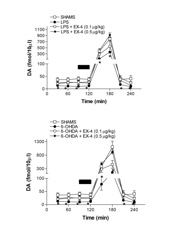
Results: EX-4 (0.1 and 0.5 mug/kg) was given seven days after intracerebral toxin injection. Seven days later circling behaviour was measured following apomorphine challenge. Circling was significantly lower in rats given EX-4 at both doses compared to animals given 6-OHDA/LPS and vehicle. Consistent with these observations, striatal tissue DA concentrations were markedly higher in 6-OHDA/LPS + EX-4 treated rats versus 6-OHDA/LPS + vehicle groups, whilst assay of L-DOPA production by tyrosine hydroxylase was greatly reduced in the striata of 6-OHDA/LPS + vehicle rats, but this was not the case in rats co-administered EX-4. Furthermore nigral TH staining recorded in 6-OHDA/LPS + vehicle treated animals was markedly lower than in sham-operated or EX-4 treated rats. Finally, EX-4 clearly reversed the loss of extracellular DA in the striata of toxin lesioned freely moving rats.

Conclusion: The apparent ability of EX-4 to arrest progression of, or even reverse nigral lesions once established, suggests that pharmacological manipulation of the GLP-1 receptor system could have substantial therapeutic utility in PD. Critically, in contrast to other peptide agents that have been demonstrated to possess neuroprotective properties in pre-clinical models of PD, EX-4 is in current clinical use in the management of type-II diabetes and freely crosses the blood brain barrier; hence, assessment of the clinical efficacy of EX-4 in patients with PD could be pursued without delay.


Figures

Figure 1
Figure 1
Flow diagram of the protocols used in the present study and the sequence in which they were performed. Groups of rats were put through all of the procedures from toxin injection to ex vivo neurochemistry and histology.
Figure 2
Figure 2
Effect of EX-4 (0.1 and 0.5 μg/kg) on apomorphine-induced rotational behaviour in 6-OHDA (upper panel) or LPS (lower panel) lesioned rats. EX-4 was administered twice daily for seven days seven days after toxin injection. Circling was measured for 120 s 30 min after apomorphine injection. One way ANOVA values were 14.90, p < 0.001 (6-OHDA) and 16.24, p < 0.001 (LPS). *indicates significant differences from UCN or sham injection sites (p < 0.01, n = 6 per group).
Figure 3
Figure 3
Effect of EX-4 (0.1 and 0.5 μg/kg) on striatal tissue DA content in 6-OHDA (upper panel) or LPS (lower panel) lesioned rats. EX-4 was administered twice daily for seven days seven days after toxin injection. One way ANOVA values were 7.88, p < 0.001 (6-OHDA) and 6.67, p < 0.001 (LPS). *indicates significant differences from EX-4 or sham injection sites (p < 0.01, n = 6 per group).
Figure 4
Figure 4
Effect of EX-4 (0.1 and 0.5 μg/kg) on striatal tissue TH activity in 6-OHDA (upper panel) or LPS (lower panel) lesioned rats. EX-4 was administered twice daily for seven days seven days after toxin injection. One way ANOVA values were 7.88, p < 0.001 (6-OHDA) and 6.67, p < 0.001 (LPS). *indicates significant differences from EX-4 or sham injection sites (p < 0.01, n = 6 per group).
Figure 5
Figure 5
Photomicrographs of selected rat nigral sections, immunostained for TH. Nigrostriatal cell bodies and dendrites appear dark brown/brown in sections. Sections A, C, E and are contralateral (untreated) nigra for comparison with ipsilateral (treated) nigra B, D, F. Key: B – 6-OHDA + vehicle, D – 6-OHDA + EX-4 0.1 μg/kg, F – 6-OHDA + EX-4 0.5 μg/kg. Bar is 100 μm. Sections are representative of 6 rats for each treatment.
Figure 6
Figure 6
Photomicrographs of selected rat nigral sections, immunostained for TH. Nigrostriatal cell bodies and dendrites appear dark brown/brown in sections. Sections A, C, E and are contralateral (untreated) nigra for comparison with ipsilateral (treated) nigra B, D, F. Key: B – LPS + vehicle, D – LPS + EX-4 0.1 μg/kg, F – LPS + EX-4 0.5 μg/kg. Bar is 100 μm. Sections are representative of 6 rats for each treatment.
Figure 7
Figure 7
Effect of EX-4 on striatal extracellular DA in 6-OHDA (upper panel) and LPS (lower panel) lesioned rats. The bar indicates the period of 100 mM K+ infusion. Two way ANOVA between treatments were F = 6.16 and 8.77 between treatments and 156.3 and 187.6 over time in 6-OHDA and LPS treated groups respectively. *Denotes differences from sham or EX-4 co-treated rats using Bonferonni's multiple comparison test post hoc. (p < 0.05, n = 6 per group).

References

    1. Clark D, White FJ. Review: D1 dopamine receptor B the search for a function: a critical evaluation of the D1/D2 dopamine receptor classification and its functional implications. Synapse. 1987;1:347–388. doi: 10.1002/syn.890010408. - DOI - PubMed
    1. Vaux DL, Korsmeyer SJ. Cell death in development. Cell. 1999;96:245–254. doi: 10.1016/S0092-8674(00)80564-4. - DOI - PubMed
    1. Gandhi S, Wood NW. Molecular pathogenesis of Parkinson's disease. Hum Mol Genet. 2005;14:2749–2755. doi: 10.1093/hmg/ddi308. - DOI - PubMed
    1. Block ML, Zecca L, Hong J-S. Microglia-mediated neurotoxicity: Uncovering the molecular mechanisms. Nature Neurosci. 2007;8:57–69. doi: 10.1038/nrn2038. - DOI - PubMed
    1. Whitton PS. Inflammation as a potential causative factor in the aetiology of Parkinsons disease. Br J Pharmacol. 2007;150:963–976. doi: 10.1038/sj.bjp.0707167. - DOI - PMC - PubMed

Publication types

MeSH terms

LinkOut - more resources