. 2008 Jun;36(11):3802-18.
doi: 10.1093/nar/gkn271. Epub 2008 May 20.

In vivo identification of novel STAT5 target genes

Affiliations

In vivo identification of novel STAT5 target genes

Beth Basham et al. Nucleic Acids Res. 2008 Jun.

Abstract

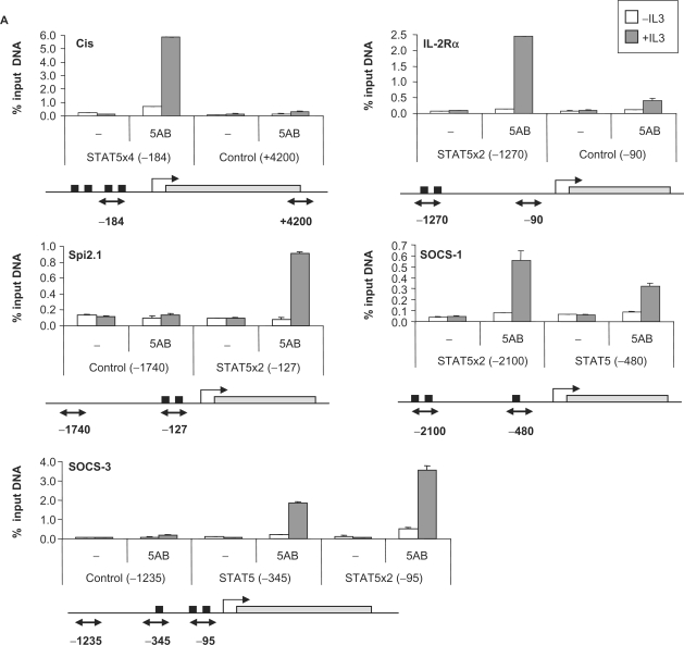
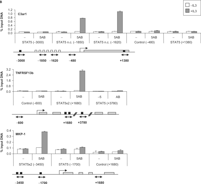
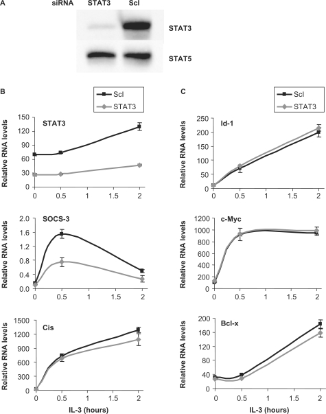
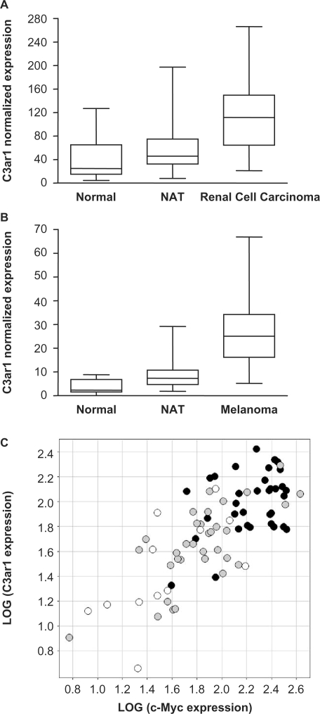
STAT5A and STAT5B proteins belong to the family of signal transducers and activators of transcription. They are encoded by two separate genes with 91% identity in their amino acid sequences. Despite their high degree of conservation, STAT5A and STAT5B exert non-redundant functions, resulting at least in part from differences in target gene activation. To better characterize the differential contribution of STAT5A and STAT5B in gene regulation, we performed single or double knockdown of STAT5A and STAT5B using small interfering RNA. Subsequent gene expression profiling and RT-qPCR analyses of IL-3-stimulated Ba/F3-beta cells led to the identification of putative novel STAT5 target genes. Chromatin immunoprecipitation assays analyzing the corresponding gene loci identified unusual STAT5 binding sites compared to conventional STAT5 responsive elements. Some of the STAT5 targets identified are upregulated in several human cancers, suggesting that they might represent potential oncogenes in STAT5-associated malignancies.


Figures

Figure 1.
Figure 1.
STAT5 mRNA levels in Ba/F3-β cells upon STAT5A and STAT5B double (A) and single (B) knockdown. Ba/F3-β cells were transfected either with a non-specific siRNA (ScI), or with siRNAs specific for STAT5A (5A), STAT5B (5B) or both (5AB). Twenty-six hours later, cells were starved for 5 h and stimulated with IL-3. Cells were harvested at the indicated times, and mRNAs levels were monitored by RT-qPCR. A 60% reduction in STAT5 mRNA levels was detected in both the single and double knockdown, with the expected specificity. Reduction in STAT5 protein levels was verified by western blot and has been already published (46). STAT3 mRNA levels (this figure), as well as protein levels (46), remained unaffected, confirming the specificity of STAT5 targeting.
Figure 2.
Figure 2.
Expression of known and putative STAT5 target genes in STAT5A and STAT5B double (A) and single (B) knockdown. RNAs were isolated from Ba/F3-β cells treated as described in Figure 1, and analyzed by RT-qPCR. Six distinct expression profiles could be recognized, represented here by selected genes (see also Table 2), revealing a clear differential contribution of STAT5A and STAT5B in regulating expression of STAT5 target genes.
Figure 3.
Figure 3.
STAT3 and STAT5 do not have redundant functions in regulating STAT5 target gene expression in IL-3-stimulated Ba/F3-β cells. Ba/F3-β cells were transfected with a siRNA specific for STAT3 or with a control siRNA (ScI), as described in Figure 1. (A) At the time of IL-3 stimulation, a specific reduction in STAT3 protein levels upon STAT3 knockdown was verified by western blot using antibodies directed against STAT3 or STAT5 as a control. (B and C) mRNA levels for STAT3, SOCS-3—a STAT3 target—and Cis, Id-1, c-Myc and Bcl-x were analyzed as described in Figures 1 and 2.
Figure 4.
Figure 4.
STAT5 recruitment in vivo on known (A) and novel (B) STAT5 target genes. Ba/F3-β cells were stimulated with IL-3 for 45 min and ChIP was performed as described in Materials and Methods section, using antibodies directed against STAT5A and STAT5B (5AB) or no antibody as a control (−). The isolated genomic DNA was analyzed by real-time PCR using primers specific for STAT5 binding sites or control regions, as indicated under each graph. Black squares indicate consensus STAT5 binding sites, open squares non-consensus (n.c.) STAT5 binding sites and gray rectangles represent open reading frames (A) or exons (B). Exons/introns were not represented in (A) for convenience. Positions are relative to the transcription start site.
Figure 4.
Figure 4.
STAT5 recruitment in vivo on known (A) and novel (B) STAT5 target genes. Ba/F3-β cells were stimulated with IL-3 for 45 min and ChIP was performed as described in Materials and Methods section, using antibodies directed against STAT5A and STAT5B (5AB) or no antibody as a control (−). The isolated genomic DNA was analyzed by real-time PCR using primers specific for STAT5 binding sites or control regions, as indicated under each graph. Black squares indicate consensus STAT5 binding sites, open squares non-consensus (n.c.) STAT5 binding sites and gray rectangles represent open reading frames (A) or exons (B). Exons/introns were not represented in (A) for convenience. Positions are relative to the transcription start site.
Figure 5.
Figure 5.
C3ar1, a novel STAT5 target gene, is overexpressed in (A, C) renal cell carcinoma and (B) melanoma. Expression of C3ar1 and c-Myc was evaluated by RT-qPCR on a panel of cDNAs samples from various human tumors and the corresponding NAT, as well as from normal tissues, as described in Materials and Methods section. mRNA levels were normalized to ubiquitin mRNA levels. C3ar1 is overexpressed in renal cell carcinomas (n = 35) (A) and melanoma tumors (n = 94) (B) compared to normal tissues [renal (n = 12) and skin (n = 15), respectively] and NAT [NAT renal (n = 33) and NAT skin (n = 91), respectively]. P-values for the difference in medians between tumors, normal tissues and NAT are <0.0001 by a two-tailed non-parametric Mann-Whitney test (Graph Pad Prism). The box represents 25–75th percentile of the data, the line represents the median and whiskers are the minimum and maximum values. (C) Expression of C3ar1 and c-Myc is upregulated in renal cell carcinoma (black dots) compared to NAT (gray dots) and normal tissues (white dots). Additionally, expression of C3ar1 and c-Myc is correlated in renal tumor and normal tissue. The squared correlation value, R2 is 55%.

References

    1. Ivashkiv LB, Hu X. Signaling by STATs. Arthritis Res. Ther. 2004;6:159–168. - PMC - PubMed
    1. Bowman T, Garcia R, Turkson J, Jove R. STATs in oncogenesis. Oncogene. 2000;19:2474–2488. - PubMed
    1. Calo V, Migliavacca M, Bazan V, Macaluso M, Buscemi M, Gebbia N, Russo A. STAT proteins: from normal control of cellular events to tumorigenesis. J. Cell. Physiol. 2003;197:157–168. - PubMed
    1. Grimley PM, Dong F, Rui H. Stat5a and Stat5b: fraternal twins of signal transduction and transcriptional activation. Cytokine Growth Factor Rev. 1999;10:131–157. - PubMed
    1. Gouilleux F, Wakao H, Mundt M, Groner B. Prolactin induces phosphorylation of Tyr694 of Stat5 (MGF), a prerequisite for DNA binding and induction of transcription. EMBO J. 1994;13:4361–4369. - PMC - PubMed

Publication types

MeSH terms

Substances