Comment
. 2008 May 27;105(21):7343-4.
doi: 10.1073/pnas.0804122105. Epub 2008 May 20.

Rhodopsin's active state is frozen like a DEER in the headlights

Affiliations
Comment

Rhodopsin's active state is frozen like a DEER in the headlights

Thomas Huber et al. Proc Natl Acad Sci U S A. .
No abstract available


Conflict of interest statement

The authors declare no conflict of interest.

Figures
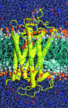
Fig. 1.
Fig. 1.
Dynamic receptors. Rhodopsin is depicted in a phospholipid bilayer membrane. The structure was simulated in a model bilayer environment (15-ns and 45,000 atoms) starting from the crystal structure (1HZX) (19). The retinylidene ligand is shown in red; the lipid acyl carbonyl carbons representing the hydrophobic boundary are shown as white balls; and the phosphates representing the headgroups are in orange. The extracellular surface is toward the top and the cytoplasmic surface toward the bottom. DEER shows that the cytoplasmic end of TM6 moves away from the center of the receptor upon activation, which likely requires rigid-body movement of the helix.

Comment on

References

    1. Cherezov V, et al. High-resolution crystal structure of an engineered human β2-adrenergic G protein-coupled receptor. Science. 2007;318:1258–1265. - PMC - PubMed
    1. Rasmussen SGF, et al. Crystal structure of the human β2 adrenergic G-protein-coupled receptor. Nature. 2007;450:383–388. - PubMed
    1. Okada T, et al. Functional role of internal water molecules in rhodopsin revealed by x-ray crystallography. Proc Natl Acad Sci USA. 2002;99:5982–5987. - PMC - PubMed
    1. Palczewski K, et al. Crystal structure of rhodopsin: A G protein-coupled receptor. Science. 2000;289:739–745. - PubMed
    1. Altenbach C, Kusnetzow AK, Ernst OP, Hofmann KP, Hubbell WL. High-resolution distance mapping in rhodopsin reveals the pattern of helix movement due to activation. Proc Natl Acad Sci USA. 2008;105:7439–7444. - PMC - PubMed

MeSH terms

LinkOut - more resources