. 2008 Aug;18(8):1282-93.
doi: 10.1101/gr.072090.107. Epub 2008 May 20.

A gene duplication affecting expression of the ovine ASIP gene is responsible for white and black sheep

Affiliations

A gene duplication affecting expression of the ovine ASIP gene is responsible for white and black sheep

Belinda J Norris et al. Genome Res. 2008 Aug.

Abstract

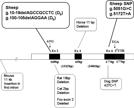
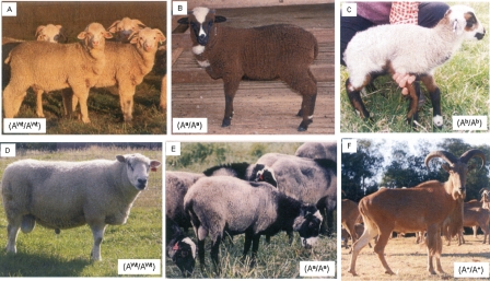
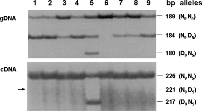
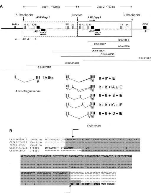
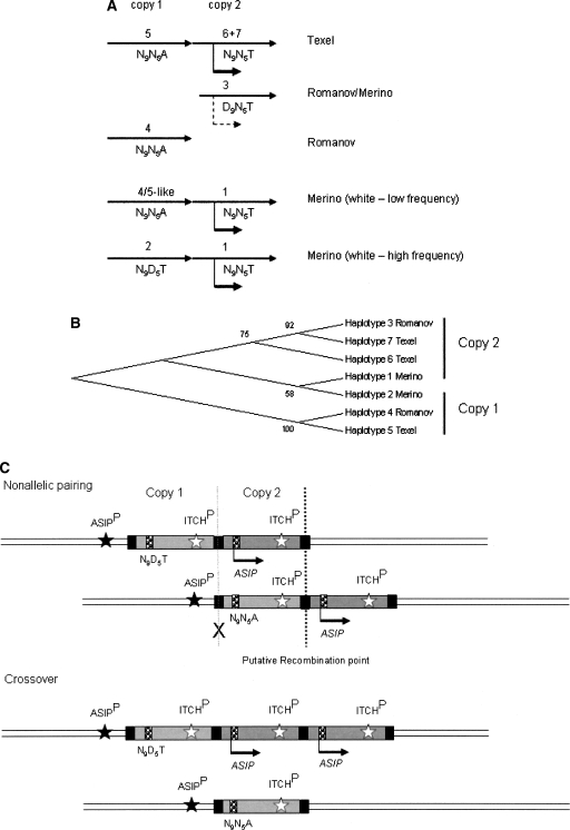
Agouti signaling protein (ASIP) functions to regulate pigmentation in mice, while its role in many other animals and in humans has not been fully determined. In this study, we identify a 190-kb tandem duplication encompassing the ovine ASIP and AHCY coding regions and the ITCH promoter region as the genetic cause of white coat color of dominant white/tan (A(Wt)) agouti sheep. The duplication 5' breakpoint is located upstream of the ASIP coding sequence. Ubiquitous expression of a second copy of the ASIP coding sequence regulated by a duplicated copy of the nearby ITCH promoter causes the white sheep phenotype. A single copy ASIP gene with a silenced ASIP promoter occurs in recessive black sheep. In contrast, a single copy functional wild-type (A(+)) ASIP is responsible for the ancient Barbary sheep coat color phenotype. The gene duplication was facilitated by homologous recombination between two non-LTR SINE sequences flanking the duplicated segment. This is the first sheep trait attributable to gene duplication and shows nonallelic homologous recombination and gene conversion events at the ovine ASIP locus could have an important role in the evolution of sheep pigmentation.


Figures

Figure 1.
Figure 1.
Schematic of the structure of the ovine ASIP gene showing the sizes of the three coding exons (black) and the intervening intron sequences (white). The exon–intron organization of the ovine gene is similar to that reported for the bovine, human, and mouse genes. Coding exons 2, 3, and 4 are separated by 1312- and 3481-bp intron sequences, respectively. The nucleotide positions of various described recessive black “non-agouti” mutations for the mouse, rat, horse, cat, fox, and dog are shown. The positions of four mutations identified from sheep in this study are shown.
Figure 2.
Figure 2.
Illustration of sheep coat color patterns. Three coat colors in the Australian Merino; (A) the dominant white; (B) the non-agouti, also known as recessive self-color-black; and (C) the badgerface pattern. The dominant white coat color in Texel is displayed in D. Romanov sheep (E) appear to have a pattern similar to recessive self-color-black and are proposed as homozygous for the most recessive ASIP allele, Aa. (F) The Barbary sheep wild-type coat color. Hypothesized genotypes for ASIP are indicated.
Figure 3.
Figure 3.
Allele-specific expression of ASIP transcripts in nine white Merino sheep. Autoradiographs of PCR products from gDNA and cDNA from the skin of nine white sheep are shown. The size difference between the 189 (226)- and 184 (221)-, and the 189 (226)- and 180 (217)-bp fragments is due to the corresponding D5 and D9 deletions in the latter fragments. Seven sheep had both the N9N5 and N9D5 alleles. In one sheep (lane 5) all three alleles N9N5, N9D5, and D9N5 were present. One sheep had only the nondeleted N9N5 alleles (lane 6). The nine white animals showed allele-specific expression of the functional N9N5 and D9N5 alleles. Expression of the nonfunctional N9D5 allele (arrow) was not detected. The different band intensities in the gDNA autoradiograph likely reflect the different allele copy numbers present in each animal.
Figure 4.
Figure 4.
RT-PCR end-point gene expression analysis of ovine ASIP and ITCH in five tissues of a white and a self-color black Merino sheep. The position of primers used to amplify each product is shown in a schematic to the right of the corresponding 1.5% agarose gel image. It, It′, and IE are noncoding exons. Open boxes Ex2, Ex3 and Ex4 (A, B, and C) and open boxes Ex17, Ex18, Ex1, and Ex2 (D, E, and F) represent coding exons of the ASIP or ITCH genes, respectively. Other alternatively spliced transcripts of the agouti gene have also amplified as evident from the bands in panel B. The most abundant ASIP transcripts are those with exon IE (∼368 bp as shown in the panel B schematic). It and It′ were both present in ITCH transcripts and generated bands of the expected size (based on similarity to the bovine sequence), but no alternatively spliced forms were evident. 3′ RACE (panel C) showed ASIP transcripts in all tissues studied to have the same polyadenylation site.
Figure 5.
Figure 5.
Schematic showing the sequenced ovine BAC clones below the structure of the ovine genomic tandem duplication and an alignment of the breakpoint and junction point sequences. (A) Noncoding exons are shown as open boxes. Protein coding exons are shown as solid black boxes. Arrows above or below the genes indicate the direction of transcription. Clones INRA-164H8, INRA-218G7, and INRA-229C6 were from the INRA Romanov Sheep BAC library (Vaiman et al. 1999), and clones CH243-160L8, CH243-234K21, CH243-455O4, CH243-373J16, and CH243-489F15 were from the CHORI-243 Texel sheep BAC library (Dalrymple et al. 2007). The three INRA BAC clones did not span the junction, 5′ or 3′ breakpoints. Clones CH243-455O4, CH243-489F15, and CH243-234K21 spanned the junction between copies; clone CH243-160L8 spanned the 3′ breakpoint and clone CH243-373J16 the 5′ breakpoint. Seven ASIP transcripts identified from the skin of a white Merino sheep (Ovis aries) and one transcript identified from the ventral skin of a Barbary sheep (Ammotragus lervia) are shown in the lower section. The coding exons 2, 3, and 4 and Barbary sheep noncoding exon 1A-like are numbered according to the nomenclature of Bultman et al. (1992) and Vrieling et al. (1994). All other noncoding exons are named alphabetically, IA to IE. Exons It and It′ are noncoding exons of both ITCH and ASIP Merino transcripts (see also Fig. 4). The positions of the 5′ and 3′ breakpoints are located 5′ of the ASIP and ITCH coding sequence regions, respectively. The ITCH promoter, including noncoding exons It, It′, and IA, was duplicated and positioned upstream of the duplicated ASIP noncoding exons IB to IE, creating a new ovine hybrid ITCH/ASIP promoter. The complete ASIP and AHCY coding exons were also within the ∼190-kb duplicated segment. Not drawn to scale. (B) DNA sequences from five Texel sheep BAC clones comprising the regions spanning the 5′ and 3′ breakpoints and the junction point between the gene duplication. Sequence identity to the master sequence is shown with a dash. The boundaries of the 143 bp of sequence similarity are marked with vertical arrows. Flanking sequences unique to the 5′ and 3′ breakpoint regions, respectively, are in bold. The highlighted (gray) regions of clone CH243-160L8 sequence of the 3′ Breakpoint was identified by Repbase (Kohany et al. 2006) as having 82% identity to a region of Bovidae non-LTR/RTE BDDF2 repetitive SINE sequence. The highlighted (gray) regions of clones CH243-373J16 (of the 5′ breakpoint) and clones CH243-489F15, CH243-234K21, and CH243-455O4 (all of the junction point) were identified as having 87%–88% identity to a region of Bovidae non-LTR BOV2 repetitive SINE sequence.
Figure 6.
Figure 6.
A model for the organization and evolution of the ovine ASIP locus in dominant white and recessive black domestic sheep. The proposed positions (A) and clustering (B) of copy 1 (inactive) and copy 2 (active) haplotypes identified from Texel, Romanov, and Merino sheep are shown. Solid arrows indicate high levels of expression of haplotypes at copy 2. A dashed arrow indicates expression of a haplotype 3-like D9 allele detected in Merino skin. The two haplotypes identified in the Romanov BAC library may not be derived from a duplicated ASIP-ITCH region as pure Romanov sheep have a recessive black-like phenotype. However, haplotype 4 clusters with haplotype 5 in copy 1 of the Texel and haplotype 3 clusters with haplotype 6 and 7 from the Texel and haplotype 1 from the Merino (B). This clustering suggests that the Romanov haplotypes are derived from the two different copies of the putative original gene duplication. The low frequency presence of a haplotype 4/5-like copy 1 region in the Merino sheep population is deduced from the identification of N9N5A genotypes in the black Merinos and is consistent with the organization of the region in the Texel sheep CHORI-243 library BACs. (C) Schematic showing resolution of nonallelic pairing between duplicated ASIP-ITCH copies with crossover products showing reciprocal deletion and triplication. The 190-kb segments of copy 1 and copy 2 of a white Merino are shown as light and dark gray boxes, respectively. Black boxes indicate the similar SINE sequence regions at the junction, 5′ and 3′ breakpoints. In this example, the ASIP genotype shown represents a white animal heterozygous at copy 1 for the nonfunctional alleles (N9D5T/N9N5A) and homozygous at copy 2 for the functional alleles (N9N5T/N9N5T). Arrows below the ASIP genes indicate the direction of transcription driven by the ITCH promoter (ITCHP). Nonallelic pairing and crossover between the junction and 3′ breakpoints (dark dashed line) would result in creation of a single copy nonfunctional (N9N5A) allele, as shown. The resulting single ASIP copy is not expressed as the ancestral ASIP promoter (ASIPP) is silent. A crossover point (gray cross) that would result in the positioning of a nonfunctional (N9N5A) allele under the regulation of the duplicated ITCH promoter is unlikely to occur as expression of “A” alleles was not detected in Merino sheep. Variable positioning of functional haplotypes at copy 1 and 2 combined with nonallelic pairing and crossover explains the different single copy alleles identified in black Merinos. Mutations and gene conversion events could also contribute to the diversity at the locus.

Similar articles

Cited by

References

    1. Adalsteinsson S. Colour inheritance in Icelandic sheep and relation between colour, fertility and fertilisation. J. Agric. Res. Iceland. 1970;2:3–135.
    1. Adalsteinsson S. Depressed fertility in Icelandic sheep caused by a single colour gene. Ann. Genet. Sel. Anim. 1975;7:445–447. - PMC - PubMed
    1. Bennett D.C., Lamoreux M.L., Lamoreux M.L. The colour loci of mice—A genetic century. Pigment Cell Res. 2003;16:333–344. - PubMed
    1. Bischof J.M., Chiang A.R., Scheetz T.E., Stone E.M., Casavant T.L., Sheffield V.C., Braun T.A., Chiang A.R., Scheetz T.E., Stone E.M., Casavant T.L., Sheffield V.C., Braun T.A., Scheetz T.E., Stone E.M., Casavant T.L., Sheffield V.C., Braun T.A., Stone E.M., Casavant T.L., Sheffield V.C., Braun T.A., Casavant T.L., Sheffield V.C., Braun T.A., Sheffield V.C., Braun T.A., Braun T.A. Genome-wide identification of pseudogenes capable of disease-causing gene conversion. Hum. Mutat. 2006;27:545–552. - PubMed
    1. Bonilla C., Boxill L.A., Donald S.A., Williams T., Sylvester N., Parra E.J., Dios S., Norton H.L., Shriver M.D., Kittles R.A., Boxill L.A., Donald S.A., Williams T., Sylvester N., Parra E.J., Dios S., Norton H.L., Shriver M.D., Kittles R.A., Donald S.A., Williams T., Sylvester N., Parra E.J., Dios S., Norton H.L., Shriver M.D., Kittles R.A., Williams T., Sylvester N., Parra E.J., Dios S., Norton H.L., Shriver M.D., Kittles R.A., Sylvester N., Parra E.J., Dios S., Norton H.L., Shriver M.D., Kittles R.A., Parra E.J., Dios S., Norton H.L., Shriver M.D., Kittles R.A., Dios S., Norton H.L., Shriver M.D., Kittles R.A., Norton H.L., Shriver M.D., Kittles R.A., Shriver M.D., Kittles R.A., Kittles R.A. The 8818G allele of the agouti signaling protein (ASIP) gene is ancestral and is associated with darker skin colour in African Americans. Hum. Genet. 2005;116:402–406. - PubMed

Publication types

MeSH terms