. 2008 May 21;3(5):e2213.
doi: 10.1371/journal.pone.0002213.

Replicative senescence of mesenchymal stem cells: a continuous and organized process

Affiliations

Replicative senescence of mesenchymal stem cells: a continuous and organized process

Wolfgang Wagner et al. PLoS One. .

Abstract

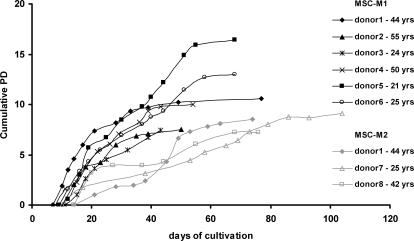
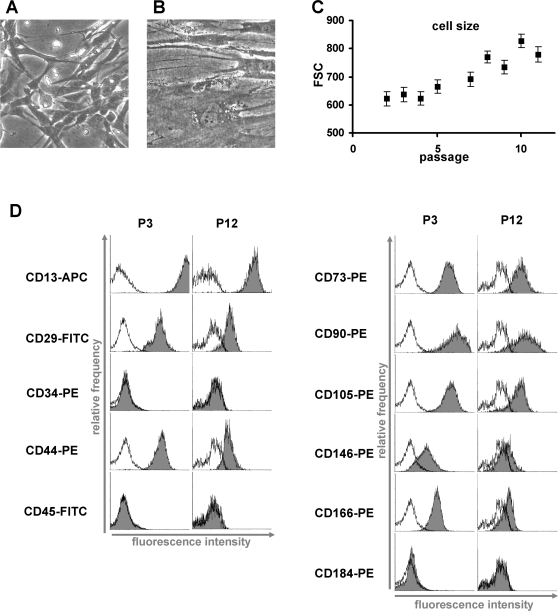
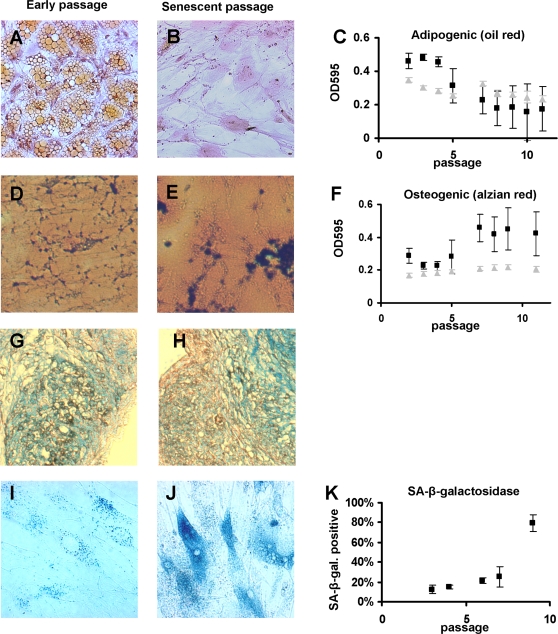
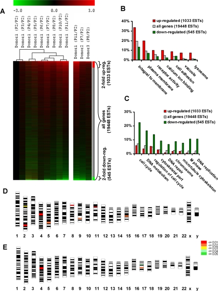
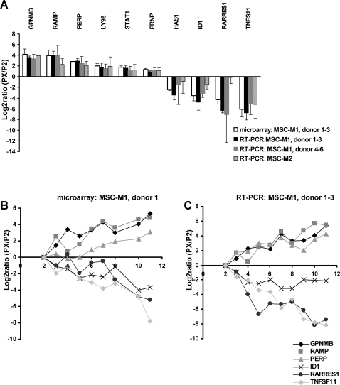
Mesenchymal stem cells (MSC) comprise a promising tool for cellular therapy. These cells are usually culture expanded prior to their application. However, a precise molecular definition of MSC and the sequel of long-term in vitro culture are yet unknown. In this study, we have addressed the impact of replicative senescence on human MSC preparations. Within 43 to 77 days of cultivation (7 to 12 passages), MSC demonstrated morphological abnormalities, enlargement, attenuated expression of specific surface markers, and ultimately proliferation arrest. Adipogenic differentiation potential decreased whereas the propensity for osteogenic differentiation increased. mRNA expression profiling revealed a consistent pattern of alterations in the global gene expression signature of MSC at different passages. These changes are not restricted to later passages, but are continuously acquired with increasing passages. Genes involved in cell cycle, DNA replication and DNA repair are significantly down-regulated in late passages. Genes from chromosome 4q21 were over-represented among differentially regulated transcripts. Differential expression of 10 genes has been verified in independent donor samples as well as in MSC that were isolated under different culture conditions. Furthermore, miRNA expression profiling revealed an up-regulation of hsa-mir-371, hsa-mir-369-5P, hsa-mir-29c, hsa-mir-499 and hsa-let-7f upon in vitro propagation. Our studies indicate that replicative senescence of MSC preparations is a continuous process starting from the first passage onwards. This process includes far reaching alterations in phenotype, differentiation potential, global gene expression patterns, and miRNA profiles that need to be considered for therapeutic application of MSC preparations.


Conflict of interest statement

Competing Interests: The authors have declared that no competing interests exist.

Figures

Figure 1
Figure 1. Long-term growth curves.
Long-term growth curves are demonstrated for six MSC preparations isolated under culture conditions M1 and for three MSC preparations isolated under culture conditions M2. Cell numbers were determined at the end of every passage and cumulative population doublings (PD) were calculated in relation to the cell numbers at the first passage. The age of each donor is provided.
Figure 2
Figure 2. Morphologic changes and immunophenotype upon senescence.
Replicative senescence is reflected by dramatic changes in morphology. Cells enlarge, generate more vacuoles and cellular debris and ultimately stop proliferation. Representative morphology of MSC in early (P3) and senescent passage (P12) is presented (A, B). The continuous increase in cell size and granularity is reflected by the increasing forward-scatter signal in flow cytometry (FSC, ±SD; C). Immunophenotypic analysis of all MSC preparations was in accordance with the literature whereby the detection level for positive markers was much higher in early passages compared to late passages (black line  =  autofluorescence; D). A representative analysis of three preparations is demonstrated.
Figure 3
Figure 3. In vitro differentiation.
MSC of different passages were simultaneously differentiated along adipogenic or osteogenic line. Fat accumulation was visualized by Oil Red-O staining. Adipogenic differentiation potential decreased in higher passages (A, B, C). In negative controls without differentiation (grey triangles) no fat accumulation was observed but the cells grew to a higher density which also resulted in higher OD. Osteogenic differentiation was visualized by van Kossa staining (not demonstrated) or Alizarin red staining. There was a higher propensity for osteogenic differentiation in higher cell passages (D, E, F). Senescence associated β-galactosidase staining increases in the later passages (G, H, I). Representative results of three independent MSC preparations are demonstrated (±SD).
Figure 4
Figure 4. mRNA expression profile of MSC changes extensively with higher passage.
Differential gene expression of senescent passages versus P2 was analyzed by Affymetix GeneChip technology in three independent MSC preparations. 19,448 ESTs that were detected as present in at least 10 of 13 hybridizations were ordered according to their log2ratio. 1033 ESTs were more than 2-fold up-regulated (red) and 545 were more than 2-fold down-regulated (green). Analysis of different passages of donor1 demonstrated increasing changes in the global gene expression pattern during in vitro senescence (A). GeneOnthology analysis was performed for the subsets of genes that were >2-fold up-regulated or >2-fold down-regulated in comparison to all genes detected as present on the microarray. The percentages of genes that contributed to representative categories are depicted (B,C; P<0.0001). Probabilities of co-localization of regulated genes plotted onto a human karyogram. The probability of representation of 2-fold up-regulated genes (D) and 2-fold down-regulated genes (E) on chromosomal regions is indicated by color coding.
Figure 5
Figure 5. QRT-PCR validation of mRNA expression.
Differential expression of senescent passage (PX) versus P2 was validated by using QRT-PCR for 10 genes (A). Results were in line with microarray data for all tested genes, investigating either the same three MSC preparations (donor 1–3) or three independent donor samples that were isolated in the same culture medium M1 (donor 4–6). Furthermore, differential gene expression was also observed in three MSC preparations isolated under different culture conditions (M2). Differential mRNA expression was not restricted to senescent passages but increased during the course of replicative senescence (B,C).
Figure 6
Figure 6. miRNA expression changes upon in vitro senescence.
miRNA expression in early and senescent passages of three MSC preparations was determined by microarray analysis (miCHIP) . Five miRNAs that are up-regulated during senescence are depicted (*  =  significant by SAM analysis). miRNA expression was also analyzed in the sequential passages of donor 1 and hierarchical cluster analysis revealed that expression of these miRNAs was overall increased during senescence (A). Furthermore, differential miRNA expression was validated by QRT-PCR for hsa-mir-29c, hsa-mir-369-5p and hsa-let-7f in the three MSC preparations that were used for microarray analysis as well as in three additional samples (B).

References

    1. Reyes M, Lund T, Lenvik T, Aguiar D, Koodie L, et al. Purification and ex vivo expansion of postnatal human marrow mesodermal progenitor cells. Blood. 2001;98:2615–2625. - PubMed
    1. Jiang Y, Jahagirdar BN, Reinhardt RL, Schwartz RE, Keene CD, et al. Pluripotency of mesenchymal stem cells derived from adult marrow. Nature. 2002;418:41–49. - PubMed
    1. Petersen BE, Bowen WC, Patrene KD, Mars WM, Sullivan AK, et al. Bone marrow as a potential source of hepatic oval cells. Science. 1999;284:1168–1170. - PubMed
    1. Schwartz RE, Reyes M, Koodie L, Jiang Y, Blackstad M, et al. Multipotent adult progenitor cells from bone marrow differentiate into functional hepatocyte-like cells. J Clin Invest. 2002;109:1291–1302. - PMC - PubMed
    1. Horwitz EM, Le BK, Dominici M, Mueller I, Slaper-Cortenbach I, et al. Clarification of the nomenclature for MSC: The International Society for Cellular Therapy position statement. Cytotherapy. 2005;7:393–395. - PubMed

Publication types