Case Reports
. 2008:2008:471512.
doi: 10.1155/2008/471512.

Successful Multimodality Endoscopic Treatment of Gastric Outlet Obstruction Caused by an Impacted Gallstone (Bouveret's Syndrome)

Affiliations
Case Reports

Successful Multimodality Endoscopic Treatment of Gastric Outlet Obstruction Caused by an Impacted Gallstone (Bouveret's Syndrome)

Jason N Rogart et al. Diagn Ther Endosc. 2008.

Abstract

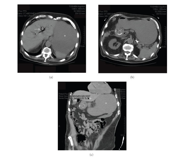
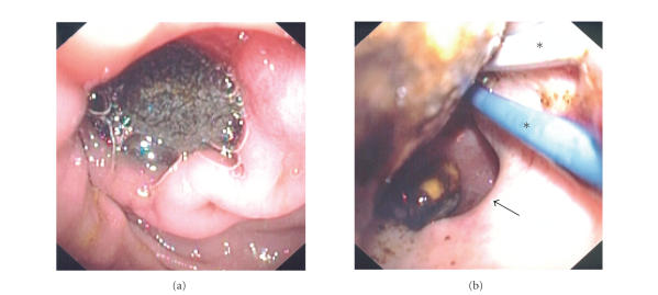
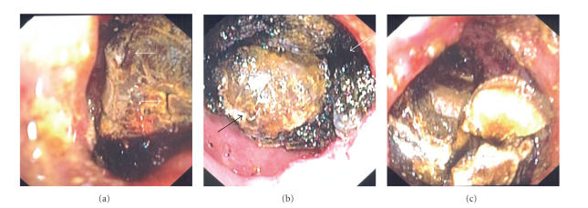
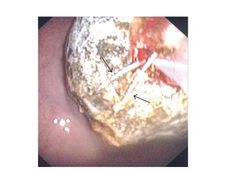
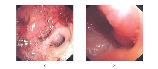
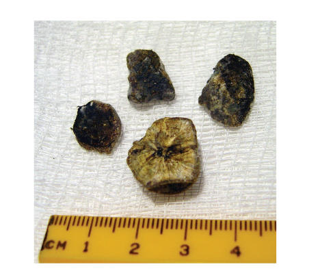
Bouveret's syndrome is a rare condition of gastric outlet obstruction resulting from the migration of a gallstone through a choledochoduodenal fistula. Due to the large size of these stones and the difficult location in which they become impacted, endoscopic treatment is unsuccessful and most patients require surgery. We report the case of an elderly male who presented with nausea and hematemesis, and was found on CT scan and endoscopy to have an obstructing gallstone in his duodenal bulb. After several endoscopic sessions and the use of multiple instruments including a Holmium: YAG laser and electrohydraulic lithotripter, fragmentation and endoscopic removal of the stone were successful. We believe this to be the first case of Bouveret's syndrome successfully treated by endoscopy alone in the United States. We describe the difficulties encountered which necessitated varied and innovative therapeutic techniques.


Figures

Figure 1
Figure 1
Computed tomography (CT) scan of abdomen and pelvis. (a) Axial image showing pneumobilia (arrow) and a dilated fluid-filled stomach (*). (b) 1-2 large gallstones (arrow) can be seen within an area of inflammation where the gallbladder is in close proximity to the duodenum. (c) Coronal reconstruction showing gallstone within duodenum (long arrow), Pneumobilia (short arrows), and dilated stomach (*) are also seen.
Figure 2
Figure 2
(a) large gallstone in the duodenal bulb, obstructing the pylorus. (b) Attempts to extract the stone failed with multiple instruments, including biliary and CRE balloons (*). The orifice of the choledochoduodenal fistula (arrow) can be seen.
Figure 3
Figure 3
Two different lithotripters were used to fragment the stone. (a) Holmium: YAG laser produced small cracks on the proximal surface (arrows) but the majority of the stone still remained impacted. (b) Electrohydraulic lithotripsy (IEHL) successfully shattered the outer “shell” of the stone (white arrow) and left behind a smaller, much harder core (black arrow). (c) Ultimately, the majority of the stone was fragmented after extensive use of both lithotriptors.
Figure 4
Figure 4
Double-snare extraction technique. Two overlapping jumbo polypectomy snares (arrows) were used to grasp the stone at different angles, providing adequate leverage for extraction into the stomach.
Figure 5
Figure 5
Visualization of the choledochoduodenal fistula. (a) After stone extraction, the large orifice of the fistula (arrow) can be seen in the duodenal bulb, whose mucosa is diffusely ulcerated. (b) The gastroscope passed easily through the fistula into the lumen of the gallbladder.
Figure 6
Figure 6
The largest stone fragments were removed perorally. The inner composition of the largest piece can be seen, measuring greater than 1 cm in diameter.

References

    1. Lowe AS, Stephenson S, Kay CL, May J. Duodenal obstruction by gallstone (Bouveret's syndrome): a review of the literature. Endoscopy. 2005;37(1):82–87. - PubMed
    1. Bouveret L. Sténose du pylore adhérent à la vésicule. Revue Médicale. 1896;16:1–16.
    1. Gencosmanoglu R, Inceoglu R, Baysal C, Akansel S, Tozun N. Bouveret's syndrome complicated by a distal gallstone ileus. World Journal of Gastroenterology. 2003;9(12):2873–2875. - PMC - PubMed
    1. Bedogni G, Contini S, Meinero M. Pyloroduodenal obstruction due to a biliary stone (Bouveret's syndrome) managed by endoscopic extraction. Gastrointestinal Endoscopy. 1985;31(1):36–38. - PubMed
    1. Moriai T, Hasegawa T, Fuzita M, Kimura A, Tani T, Makino I. Successful removal of massive intragastric gallstones by endoscopic electrohydraulic lithotripsy and mechanical lithotripsy. American Journal of Gastroenterology. 1991;86(5):627–629. - PubMed

Publication types

LinkOut - more resources