. 2008 May 21;3(5):e2201.
doi: 10.1371/journal.pone.0002201.

Adaptive evolution and functional redesign of core metabolic proteins in snakes

Affiliations

Adaptive evolution and functional redesign of core metabolic proteins in snakes

Todd A Castoe et al. PLoS One. .

Abstract

Background: Adaptive evolutionary episodes in core metabolic proteins are uncommon, and are even more rarely linked to major macroevolutionary shifts.

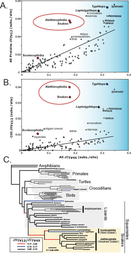
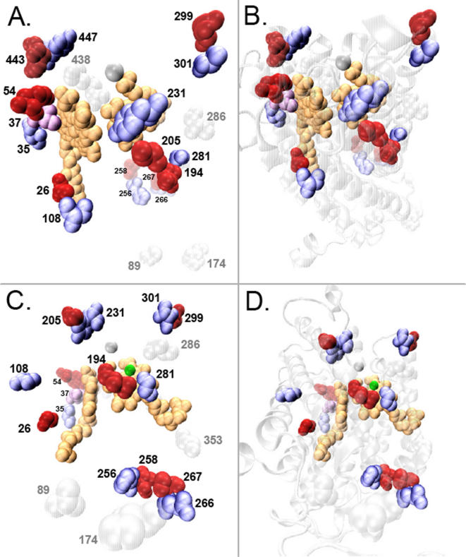
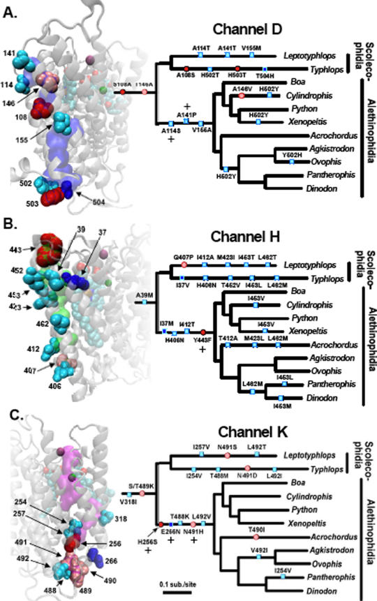
Methodology/principal findings: We conducted extensive molecular evolutionary analyses on snake mitochondrial proteins and discovered multiple lines of evidence suggesting that the proteins at the core of aerobic metabolism in snakes have undergone remarkably large episodic bursts of adaptive change. We show that snake mitochondrial proteins experienced unprecedented levels of positive selection, coevolution, convergence, and reversion at functionally critical residues. We examined Cytochrome C oxidase subunit I (COI) in detail, and show that it experienced extensive modification of normally conserved residues involved in proton transport and delivery of electrons and oxygen. Thus, adaptive changes likely altered the flow of protons and other aspects of function in CO, thereby influencing fundamental characteristics of aerobic metabolism. We refer to these processes as "evolutionary redesign" because of the magnitude of the episodic bursts and the degree to which they affected core functional residues.

Conclusions/significance: The evolutionary redesign of snake COI coincided with adaptive bursts in other mitochondrial proteins and substantial changes in mitochondrial genome structure. It also generally coincided with or preceded major shifts in ecological niche and the evolution of extensive physiological adaptations related to lung reduction, large prey consumption, and venom evolution. The parallel timing of these major evolutionary events suggests that evolutionary redesign of metabolic and mitochondrial function may be related to, or underlie, the extreme changes in physiological and metabolic efficiency, flexibility, and innovation observed in snake evolution.


Conflict of interest statement

Competing Interests: The authors have declared that no competing interests exist.

Figures

Figure 1
Figure 1. Mitochondrial proteins have experienced extraordinarily elevated rates of amino acid replacement early in the evolution of snakes.
The conservative transversion-based approximations of the relative rates of non-synonymous to synonymous substitution (dN TV12 / dS TV4x) rates are shown as open or colored circles for each branch of the phylogenetic tree; linear regression lines (excluding points in the red ellipse) are shown in black (A and B). The calculations shown are from (A) all mitochondrial proteins and (B) cytochrome C oxidase subunit 1 (COI). Blue-shaded areas of A and B indicate very long branches with high dS TV4x values where the (dN TV12 / dS TV4x) estimate may be inaccurate, possibly due to dS TV4x saturation and underestimation. Note that early snake branches have very high dN TV12, far greater than branches of comparable length (dS TV4x). This is strong evidence for extraordinarily accelerated rates of amino acid replacement early in snake evolution. The phylogenetic tree of relationships among species in our comparative dataset is shown in (C). Branches with extremely high values of dN TV12 / dS TV4X for COI are indicated with colored lines (black, blue, red) following the key in the bottom left. The circles for branches in (A) and (B) were colored according to the same legend for ratios of COI (dN TV12 / dS TV4x).
Figure 2
Figure 2. Unique amino acid replacements in Cytochrome C oxidase subunit 1 form tight spatial clusters.
The twenty-three unique amino acid replacements in the cytochrome C oxidase subunit 1 (COI) protein of snakes form seven pairs and one triplet of spatially clustered amino acid replacements, concentrated at the core functional region of the COI protein. The seven spatially adjacent pairs of amino acid residues, strongly suggestive of coevolutionary adaptive change, are shown in blue/red paired spacefill combinations, and one triplet cluster is shown in a blue/purple/red combination. Unique sites that did not form clusters are shown in gray spacefill representations. The two heme groups are shown in gold spacefill shapes, the COI backbone in white, and the magnesium and copper atoms are shown as magenta and green balls, respectively. Two different perspectives are depicted, one in A and B, and a second in C and D; Figure sets A/B and C/D are the same views with B and D showing the ribbon structure of the COI backbone in transparent grey.
Figure 3
Figure 3. Amino acid replacements at residues within and directly adjacent to the three proton transport channels (D, H, and K) within cytochrome C oxidase subunit 1 (COI) have substantially altered COI structure and presumably function in snakes.
On the small snake phylogenetic trees shown, amino acid replacements that occur in snakes at sites comprising the channel are indicated with ovals, and changes at sites adjacent to each channel are indicated with rectangles. Deep colors (dark red and dark blue) indicate replacements at unique sites (see text) whereas lighter colors (light red and light blue) are not unique sites. Sites that were inferred to be under significant positive selection along the Alethinophidian snake branch (by PAML branch-site analyses) are indicated with a plus sign. The relative order of replacements along each single branch has no meaning. These same sites are shown as spacefill representations in the structure, and are labeled and shaded with the same color code as the mark on the tree. One channel is shown per subfigure (A = channel D, B = channel H, and C = channel K). The three proton channels D, H, and K are shown in transparent purple, green, or magenta volumetric representation, respectively. The entire CO1 structure is shown as a transparent grey ribbon structure, heme groups are shown as spacefill representations colored by element, and the magnesium and copper atoms are colored magenta and green, respectively.

References

    1. Stewart C-B, Schilling JW, Wilson AC. Adaptive evolution in the stomach lysozymes of foregut fermenters. Nature. 1987;330:401–404. - PubMed
    1. Chimpanzee Sequencing and Analysis Consortium. Initial sequence of the chimpanzee genome and comparison with the human genome. Nature. 2005;437:69–87. - PubMed
    1. Mikkelsen TS, Wakefield MJ, Aken B, Amemiya CT, Chang JL, et al. Genome of the marsupial Monodelphis domestica reveals innovation in non-coding sequences. Nature. 2007;447:167–177. - PubMed
    1. Dong S, Kumazawa Y. Complete mitochondrial DNA sequences of six snakes: Phylogenetic relationships and molecular evolution of genomic features. J Mol Evol. 2005;61:12–22. - PubMed
    1. Jiang ZJ, Castoe TA, Austin CC, Burbrink FT, Herron MD, et al. Comparative mitochondrial genomics of snakes: extraordinary substitution rate dynamics and functionality of the duplicate control region. BMC Evol Biol. 2007;7 - PMC - PubMed

Publication types