. 2008 Jun;4(6):562-70.
doi: 10.1039/b720018h. Epub 2008 Mar 6.

Visualizing the spatial distribution of secondary metabolites produced by marine cyanobacteria and sponges via MALDI-TOF imaging

Affiliations

Visualizing the spatial distribution of secondary metabolites produced by marine cyanobacteria and sponges via MALDI-TOF imaging

Eduardo Esquenazi et al. Mol Biosyst. 2008 Jun.

Abstract

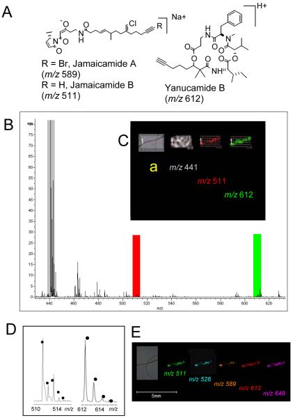
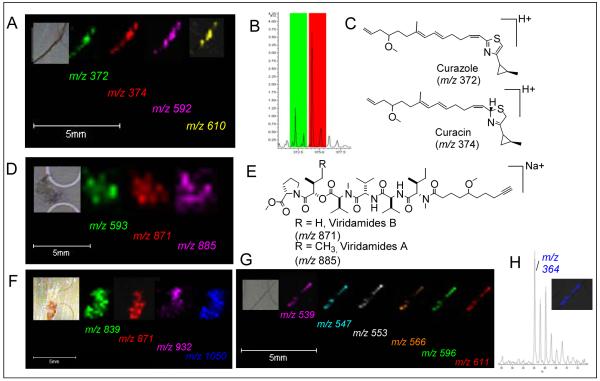
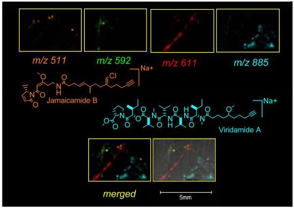
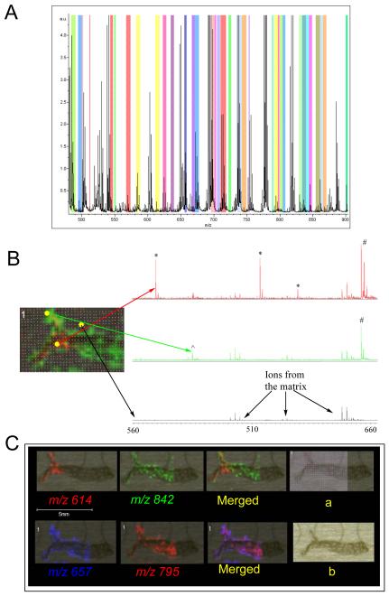
Marine cyanobacteria and sponges are prolific sources of natural products with therapeutic applications. In this paper we introduce a mass spectrometry based approach to characterize the spatial distribution of these natural products from intact organisms of differing complexities. The natural product MALDI-TOF-imaging (npMALDI-I) approach readily identified a number of metabolites from the cyanobacteria Lyngbya majuscula 3L and JHB, Oscillatoria nigro-viridis, Lyngbya bouillonii, and a Phormidium species, even when they were present as mixtures. For example, jamaicamide B, a well established natural product from the cyanobacterium Lyngbya majuscula JHB, was readily detected as were the ions that correspond to the natural products curacin A and curazole from Lyngbya majuscula 3L. In addition to these known natural products, a large number of unknown ions co-localized with the different cyanobacteria, providing an indication that this method can be used for dereplication and drug discovery strategies. Finally, npMALDI-I was used to observe the secondary metabolites found within the sponge Dysidea herbacea. From these sponge data, more than 40 ions were shown to be co-localized, many of which were halogenated. The npMALDI-I data on the sponge indicates that, based on the differential distribution of secondary metabolites, sponges have differential chemical micro-environments within their tissues. Our data demonstrate that npMALDI-I can be used to provide spatial distribution of natural products, from single strands of cyanobacteria to the very complex marine assemblage of a sponge.


Figures

Fig. 1
Fig. 1
MALDI-TOF-imaging of the intact marine cyanobacterium Lyngbya majuscula JHB filament. A) The molecular structures of jamaicamide A, B and yanucamide B. B) The average mass spectrum of a 0.6 *1.5 mm area of the MALDI imaging experiment. The colors indicate the regions visualized in C. C) The differential localization of the indicated masses with respect to the cyanobacterial filament. A shows the raster points in this experiment. D) Comparison of the theoretical isotopic distribution of jamaicamide B and yanucamide B indicated by the black dots with the observed average spectrum in this experiment. E) The spatial distribution for several molecular ions co-localized with Lyngbya majuscula JHB.
Fig. 2
Fig. 2
The spatial distribution of selected ions observed to co-localize with Lyngbya majuscula 3L (A), Oscillatoria nigro-viridis (D), and a Phormidium species (F), Lyngbya bouillonii (G). The average mass spectral trace showing curacin and curazole and the respective colors indicated is shown in figure B. The structures of curacin, curazole (C) and viridamides (E) are also shown. H) The isotopic distribution for the 364 m/z molecular ion.
Fig. 3
Fig. 3
npMALDI-I of a complex mixture of cyanobacteria: a single npMALDI-I run on a mixture of Lyngbya majuscula JHB (orange),and 3L (green), Lynbya bouillonii (Red) Oscillatoria nigro-viridis (Blue). The top panels represent detection of two known masses-Jamaicamide B (orange-structure shown) and viridamide A (blue- structure shown) as well as two unknown masses (red and green), each specifically and differentially locates to a particular organism. The bottom panels emphasizes the scale and spatial resolution, as well as the ability to visualize various different secondary metabolites from multiple organisms.
Fig. 4
Fig. 4
npMALDI-I on the sponge Dysidea herbacea. A) The average mass spectrum. The colors indicate all the ions that specifically localized to the sponge section, the colors themselves have no meaning other than that they are a means to show the differential localization. B) An image of the Dysidea section with the laser raster points and selected masses shown. In this image we show the relative ion intensities in the region from 560 to 660 m/z for three different areas of the sampling area. This image shows the different ion localizations and ion clusters associated with the matrix. Ions indicated with a # are co-localized throughout the sponge, ions that are localized near the edges of the sponge are indicated with a ^ and ions found on the inner section of the sponge are shown with a *. C) Some representative differential localizations and ion masses associated with the sponge, suggesting a differential chemical microenvironments. “a” shows the raster on the image. “b” shows the photomicrograph of the sponge-section itself.

References

    1. Newman DJ, Cragg GM. Natural Products as Sources of New Drugs over the Last 25 Years. Journal of Natural Products. 2007;70(3):461–477. - PubMed
    1. Bode BH, Mueller R. The impact of bacterial genomics on natural product research. Angewandte Chemie, International Edition. 2005;44(42):6828–6846. - PubMed
    1. Dubois J, Guenard D, Gueritte F. Recent developments in antitumour taxoids. Expert Opinion on Therapeutic Patents. 2003;13(12):1809–1823.
    1. Demain AL. Pharmaceutically active secondary metabolites of microorganisms. Applied.Microbiology and Biotechnology. 1999;52:455–463. - PubMed
    1. Taylor MW, Radax R, Steger D, Wagner M. Sponge-Associated Microorganisms: Evolution, Ecology,and Biotechnological Potential. Microbiology and Molecular Reviews. 2007;71(2):295–347. - PMC - PubMed

Publication types

MeSH terms

Substances