. 2008 Jun;12(3):914-27.
doi: 10.1111/j.1582-4934.2008.00159.x.

S100B induces tau protein hyperphosphorylation via Dickopff-1 up-regulation and disrupts the Wnt pathway in human neural stem cells

Affiliations

S100B induces tau protein hyperphosphorylation via Dickopff-1 up-regulation and disrupts the Wnt pathway in human neural stem cells

Giuseppe Esposito et al. J Cell Mol Med. 2008 Jun.

Abstract

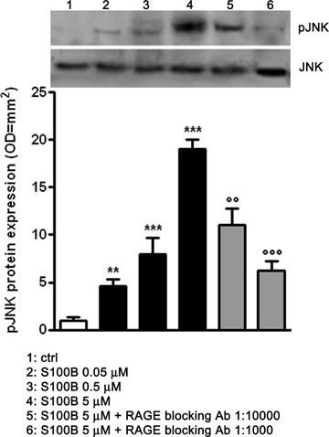
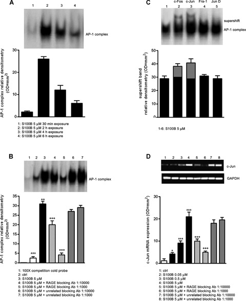
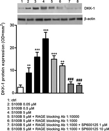
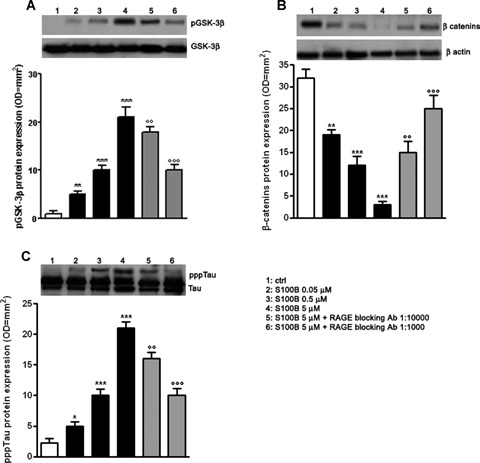
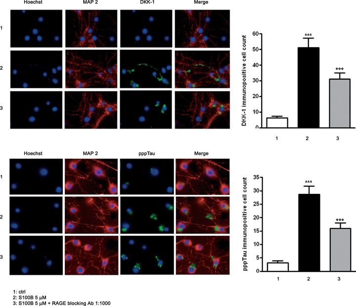
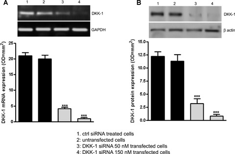
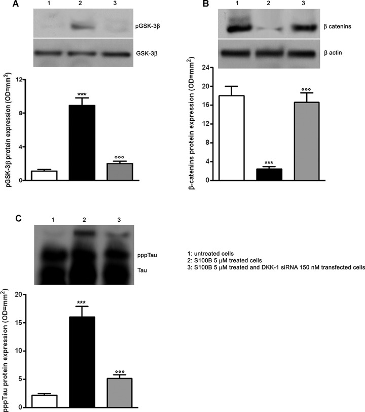
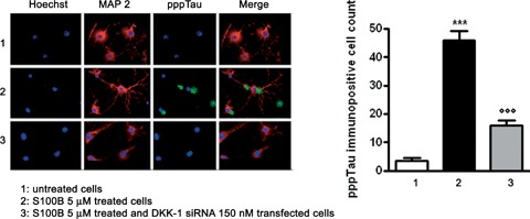
Previous studies suggest that levels of the astrocyte-derived S100B protein, such as those occurring in brain extra-cellular spaces consequent to persistent astroglial activation, may have a pathogenetic role in Alzheimer's disease (AD). Although S100B was reported to promote beta amyloid precursor protein overexpression, no clear mechanistic relationship between S100B and formation of neurofibrillary tangles (NFTs) is established. This in vitro study has been aimed at investigating whether S100B is able to disrupt Wnt pathway and lead to tau protein hyperphosphorylation. Utilizing Western blot, electrophoretic mobility shift assay, supershift and reverse transcriptase-polymerase chain reaction techniques, it has been demonstrated that micromolar S100B concentrations stimulate c-Jun N-terminal kinase (JNK) phosphorylation through the receptor for advanced glycation ending products, and subsequently activate nuclear AP-1/cJun transcription, in cultured human neural stem cells. In addition, as revealed by Western blot, small interfering RNA and immunofluorescence analysis, S100B-induced JNK activation increased expression of Dickopff-1 that, in turn, promoted glycogen synthase kinase 3beta phosphorylation and beta-catenin degradation, causing canonical Wnt pathway disruption and tau protein hyperphosphorylation. These findings propose a previously unrecognized link between S100B and tau hyperphosphorylation, suggesting S100B can contribute to NFT formation in AD and in all other conditions in which neuroinflammation may have a crucial role.


Figures

1
1
S100B induces JNK phopsphorylation through RAGE interaction in a dose-dependent fashion. pJNK protein expression was evaluated in lysates of NSCs 30 min. following exposure at increasing concentrations of S100B (0.05–5 μM), by Western blot (upper panel) and densitometric analysis of corresponding bands (lower panel). Statistics show a significant dose-dependent effect of S100B on JNK phosphorilation. Different concentrations of RAGE blocking antibody (1:1000 or 1:10,000) reverted the effect induced by the highest concentration of S100B. Results are the mean ±S.E.M. of three independent experiments. ***P < 0.001 and **P < 0.01 versus unchallenged cells (ctrl);°°°P < 0.001 and °°P < 0.01 versus 5 μM S100B challenged cells.
2
2
S100B induces AP-1/DNA complex activation through RAGE interaction. (A) AP-1/DNA complex activation was detected in NSCs at various time point (30 min. to 6 hrs) following the exposure to 5 μM of S100B. The time course of AP-1/DNA binding activity was evaluated by EMSA (upper panel) and densitometric analysis of corresponding bands (lower panel). (B) AP-1/DNA binding activity induced by 5 μM of S100B in NSCs in the presence or absence of two different concentrations of RAGE blocking antibody or unrelated blocking antibody (1:1000 or 1:10,000). Statistics show that S100B significantly enhanced the activation of AP-1/DNA complex and that the specific RAGE blocking antibody significantly blunted this effect, while the unrelated antibody failed to influence it. The AP-1/DNA binding activity was measured 2 hrs after S100B exposure by EMSA analysis (upper panel). The lower panel shows densitometric analysis of corresponding bands. (C) AP-1 antibodies (anti-Fra-1, anti-c-Fos, anti-c-Jun and anti-Jun-D) were preincubated with nuclear extracts from NSCs exposed to 5 μM of S100B. Results of supershift analysis (upper panel) demonstrate the effect of the antibodies on the changes in the relative mobility of AP-1 species 2 hrs following NSC stimulation with S100B. Densitometric analysis of corresponding bands is reported in the lower panel. (D) NSCs were challenged with increasing concentrations of S100B (0.05–5 μM) and lysed 6 hrs later. c-Jun mRNA expression was evaluated by RT-PCR (upper panel) and densitometric analysis of corresponding bands (lower panel). Statistics demonstrate significant and concentration-dependent effect of S100B on c-Jun mRNA expression. The figure also shows two different dilutions of RAGE blocking antibody (1:1000 or 1:10,000) were able to revert the effect of the highest concentration of S100B, whereas the same dilutions of an unrelated blocking antibody were ineffective. Results are the mean ±S.E.M. of three independent experiments.***P < 0.001, **P < 0.01, and *P < 0.05 versus unchallenged cells (ctrl);°°°P < 0.001 and °°P < 0.01 versus 5 μM S100B challenged cells.
3
3
S100B induces DKK-1 protein expression through RAGE and JNK involvement in a dose-dependent fashion. NSCs were challenged with increasing concentrations of S100B (0.05–5 μM) and 12 hrs later they were lysed and DKK-1 protein expression was evaluated by Western blot (upper panel) and densitometric analysis of corresponding bands (lower panel). β-actin served as a loading control. Statistics indicate that S100B significantly promoted DKK-1 protein expression in a dose-dependent fashion. Moreover two different dilutions of RAGE blocking antibody (1:1000 or 1:10,000) were able to revert in a dose-dependent manner the effect of the highest concentration of S100B. This effect was strengthened by the specific JNK phosphorylation inhibitor SP600125 (1 or 10 μM). Results are the mean ± S.E.M. of three independent experiments. ***P < 0.001 and **P < 0.01 versus unchallenged cells (ctrl);°°°P < 0.001 and °°P < 0.01 versus 5 μM S100B challenged cells; ###P < 0.001 versus 5 μM S100B plus RAGE blocking antibody (1:1000) challenged cells.
4
4
S100B induces tau protein hyperphosphorylation through Wnt pathway disruption. (A) NSCs were challenged with increasing concentrations of S100B (0.05–5 μM). The pGSK-3β protein expression was evaluated 24 hrs following treatment by Western blot (upper panel) and densitometric analysis of corresponding bands (lower panel). GSK-3β served as a loading control. Statistics demonstrate significant and concentration-dependent effect of S100B on pGSK-3β protein expression. Two different dilutions of RAGE blocking antibody (1:1000 or 1:10,000) were able to concentration-dependently antagonize the effect of the highest concentration of S100B. (B) β-catenin protein expression was evaluated 24 hrs following treatment by Western blot (upper panel) and densitometric analysis of corresponding bands (lower panel). β-actin served as a loading control. Statistics show that S100B significantly and concentration-dependently affected β-catenin protein expression. Two different dilutions of RAGE blocking antibody (1:1000 or 1:10,000) were able to revert the effect of the highest concentration of S100B in a concentration-dependent manner. (C) pppTau protein expression was evaluated 48 hrs following treatment by Western blot (upper panel) and densitometric analysis of corresponding bands (lower panel). Tau served as a loading control. Statistics indicate that S100B was able to concentration-dependently and significantly promote pppTau protein expression. Two different dilutions of RAGE blocking antibody (1:1000 or 1:10,000) antagonized the effect of the highest concentration of S100B in a concentration-dependent fashion. Results are the mean ±S.E.M. of three separated experiments.***P < 0.001, **P < 0.01, and *P < 0.05 versus unchallenged cells (ctrl);°°°P < 0.001 and °°P < 0.01 versus 5 μM S100B challenged cells.
5
5
S100B induces DKK-1 expression and tau protein hyperphosphorylation through RAGE interaction. (A) Left panel: representative photomicrographs of NSCs showing DKK-1 immunostaining (green). NSC cultures were exposed to S100B 5 μM in the presence or absence of RAGE blocking antibody (1:1000), and DKK-1 protein expression was evaluated 12 hrs later by immunofluorescence analysis. Right panel: quantification of immunoreactivity expressed as the number of DKK-1 immunopositive neuronal cells. Statistics indicate that RAGE blocking antibody significantly reverted the effect of S100B on DKK-1 protein expression. (B) Left panel: representative photomicrographs of NSCs showing pppTau immunostaining (green). NSC cultures were exposed to S100B 5 μM in the presence or absence of RAGE blocking antibody (1:1000), and pppTau protein expression was evaluated 48 hrs later by immunofluorescence analysis. Right panel: quantification of immunoreactivity expressed as the number of pppTau immunopositive neuronal cells. Statistics indicate that RAGE blocking antibody significantly reverted the effect of S100B on pppTau protein expression. Anti-MAP-2 antibody was used as a neuronal marker (red), and nuclei were stained with Hoechst 33258 (blue). Scale bar = 20 μm. Data are shown as mean ± S.E.M. of five experiments.***P < 0.001 versus unchallenged cells (ctrl);°°°P < 0.001 versus 5 μM S100B challenged cells.
6
6
DKK-1 siRNA in SHSY5Y cell cultures.(A) SHSY5Y cells were transfected with DKK-1 siRNA (50 or 150 nM) and 12 hrs later DKK-1 mRNA knockdown was evaluated by RT-PCR (upper panel). Lower panel reports the densito-metric analysis of corresponding bands. GAPDH served as a loading control.(B) DKK-1 protein expression was evaluated 24 hrs following transfection by Western blot (upper panel). In the lower panel densitometric analysis of corresponding bands is reported. β-actin served as a loading control. Untransfected and control siRNA treated cells were used as internal controls to verify the specificity of the treatment. Statistics demonstrate that DKK-1 siRNA significantly and dose-dependently induced DKK-1 mRNA knockdown in SHSY5Y cells. Results are the mean ± S.E.M. of three separated experiments.***P < 0.001 versus internal controls.
7
7
S100B requires DKK-1 activation to induce Wnt pathway disruption and tau protein hyperphosphorylation. (A) SHSY5Y cells were transfected with 150 nM DKK-1 siRNA and treated with S100B 5 μM. The pGSK-3β protein expression was evaluated 24 hrs following treatment by Western blot (upper panel) and densitometric analysis of corresponding bands (lower panel). GSK-3β served as a loading control.(B) β-catenin protein expression was evaluated 24 hrs following treatment by Western blot (upper panel) and densitometric analysis of corresponding bands (lower panel). β-actin served as a loading control.(C) pppTau protein expression was evaluated 48 hrs following treatment by Western blot (upper panel) and densitometric analysis of corresponding bands (lower panel). Tau served as a loading control. Statistics show that S100B required DKK-1 activation to reduce β-catenin protein expression and to promote pGSK-3β and pppTau protein expression. Untransfected and untreated cells were used as internal controls. Results are the mean ± S.E.M. of three independent experiments. ***P < 0.001 versus untreated cells; °°°P < 0.001 versus untrans-fected cells treated with S100B 5 μM.
8
8
DKK-1 siRNA abolishes S100B-induced tau protein hyperphosphorylation. SHSY5Y cells were transfected with 150 nM DKK-1 siRNA and treated with S100B 5 μM. pppTau protein expression was evaluated 48 hrs later by immuno-fluorescence analysis (left panel) and subsequent immunopositive cell count (right panel). Anti-MAP-2 antibody was used as a neuronal marker (red), while nuclei were stained with Hoechst 33258 (blue). Scale bar = 20 μm. Untransfected and untreated cells were used as internal controls. Results are the mean ±S.E.M. of three independent experiments.***P < 0.001 versus untreated cells;°°°P < 0.001 versus untransfected cells treated with S100B 5 μM.

References

    1. Lee VM, Balin BJ, Otvos L, Jr, Trojanowski JQ. A68: a major subunit of paired helical filaments and derivatized forms of normal tau. Science. 1991;251:675–8. - PubMed
    1. Kosik KS, Joachim CL, Selkoe DJ. Microtubule-associated protein tau (tau) is a major antigenic component of paired helical filaments in Alzheimer disease. Proc Natl Acad Sci USA. 1986;83:4044–8. - PMC - PubMed
    1. Grundke-Iqbal I, Iqbal K, Tung YC, Quinlan M, Wisniewski HM, Binder LI. Abnormal phosphorylation of the microtubule-associated protein tau (tau) in Alzheimer cytoskeletal pathology. Proc Natl Acad Sci USA. 1986;83:4913–7. - PMC - PubMed
    1. Doble BW, Woodgett JR. GSK-3: tricks of the trade for a multi-tasking kinase. J Cell Sci. 2003;116:1175–86. - PMC - PubMed
    1. De Ferrari GV, Inestrosa NC. Wnt signaling function in Alzheimer's disease. Brain Res Brain Res Rev. 2000;33:1–12. - PubMed

Publication types

MeSH terms