Review
. 2008 Aug 17;60(11):1226-1240.
doi: 10.1016/j.addr.2008.03.015. Epub 2008 Apr 10.

Bioconjugated quantum dots for in vivo molecular and cellular imaging

Affiliations
Review

Bioconjugated quantum dots for in vivo molecular and cellular imaging

Andrew M Smith et al. Adv Drug Deliv Rev. .

Abstract

Semiconductor quantum dots (QDs) are tiny light-emitting particles on the nanometer scale, and are emerging as a new class of fluorescent labels for biology and medicine. In comparison with organic dyes and fluorescent proteins, they have unique optical and electronic properties, with size-tunable light emission, superior signal brightness, resistance to photobleaching, and broad absorption spectra for simultaneous excitation of multiple fluorescence colors. QDs also provide a versatile nanoscale scaffold for designing multifunctional nanoparticles with both imaging and therapeutic functions. When linked with targeting ligands such as antibodies, peptides or small molecules, QDs can be used to target tumor biomarkers as well as tumor vasculatures with high affinity and specificity. Here we discuss the synthesis and development of state-of-the-art QD probes and their use for molecular and cellular imaging. We also examine key issues for in vivo imaging and therapy, such as nanoparticle biodistribution, pharmacokinetics, and toxicology.


Figures

Figure 1
Figure 1
Size-dependent optical properties of cadmium selenide QDs dispersed in chloroform, illustrating quantum confinement and size tunable fluorescence emission. (a) Fluorescence image of four vials of monodisperse QDs with sizes ranging from 2.2 nm to 7.3 nm in diameter. This image was obtained with ultraviolet illumination. (b) Fluorescence spectra of the same four QD samples. Narrow emission bands (23–26 nm FWHM or full-width at half-maximum) indicate narrow particle size distributions. (c) Absorption spectra of the same four QD samples. Notice that the absorption spectra are very broad, allowing a broad wavelength range for excitation. Both the absorption and emission intensities are plotted in arbitrary units (AU).
Figure 2
Figure 2
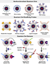
Schematic diagrams of nonfunctionalized and bioconjugated QD probes for imaging and sensing applications. See text for detailed discussion.
Figure 3
Figure 3
Encapsulation and solubilization of core-shell CdSe/CdS/ZnS quantum dots by using multivalent and hyperbranched copolymer ligands. (a) and (b) Chemical structures of PEI and 19 PEI-g-PEG copolymers consisting of two or four PEG chains per PEI polymer molecule. (c) Schematic diagram showing direct exchange reactions between the monovalent capping ligand octadecylamine and the multivalent copolymer ligands. Adapted with permission from Duan and Nie [97].
Figure 4
Figure 4
Schematic diagram showing QD interactions with blood immune cells and plasma proteins. The probable modes of interactions include (a) QD opsonization and phagocytosis by leucocytes (e.g., monocytes), (b) non-specific QD-cell membrane interactions (electrostatic or hydrophobic), and (c) fluid-phase pinocytosis.
Figure 5
Figure 5
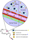
Schematic diagram showing QDs involved in both active and passive tumor targeting. In the passive mode, nanometer-sized particles such as quantum dots accumulate at tumor sites through an enhanced permeability and retention (EPR) effect [–129]. For active tumor targeting, nanoparticles are conjugated to molecular ligands such as antibodies and peptides to recognize protein targets that are over-expressed on the surface of tumor cells such as the epidermal growth factor receptor (EGFR), the transferrin receptor, or the folate receptor. Courtesy of Dr. Ximei Qian, Emory University, Atlanta, GA 30322, USA.
Figure 6
Figure 6
Schematic illustration of QD-Aptamer-Dox FRET system and its targeted delivery through receptor-mediated endocytosis. (a) QDs-aptamer conjugates (QD-Apt) are fluorescent until they are mixed with the fluorescent drug doxorubicin (Dox), which intercalates with the base pairs of the aptamer and quenches the QD via FRET. (b) Aptamer-specific endocytosis results in cellular internalization of the conjugate, release of cytotoxic Dox, and restoration of fluorescence. Adapted with permission from Bagalkof et al. [165].

Similar articles

Cited by

References

    1. Alivisatos AP. The use of nanocrystals in biological detection. Nat. Biotechnol. 2004;22:47–52. - PubMed
    1. Ferrari M. Cancer nanotechnology: Opportunities and challenges. Nat. Rev. Cancer. 2005;5:161–171. - PubMed
    1. Niemeyer CM. Nanoparticles, proteins, and nucleic acids: Biotechnology meets materials science. Angew. Chem., Int. Ed. 2001;40:4128–4158. - PubMed
    1. Cao YWC, Jin RC, Mirkin CA. Nanoparticles with Raman spectroscopic fingerprints for DNA and RNA detection. Science. 2002;297:1536–1540. - PubMed
    1. Gao XH, Yang LL, Petros JA, Marshal FF, Simons JW, Nie SM. In vivo molecular and cellular imaging with quantum dots. Curr. Opin. Biotechnol. 2005;16:63–72. - PubMed

Publication types

Substances