. 2008 Jul 25;283(30):21267-77.
doi: 10.1074/jbc.M800727200. Epub 2008 May 21.

A high affinity hepatocyte growth factor-binding site in the immunoglobulin-like region of Met

Affiliations

A high affinity hepatocyte growth factor-binding site in the immunoglobulin-like region of Met

Cristina Basilico et al. J Biol Chem. .

Abstract

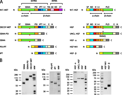
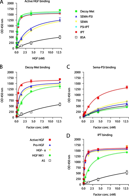
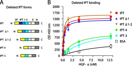
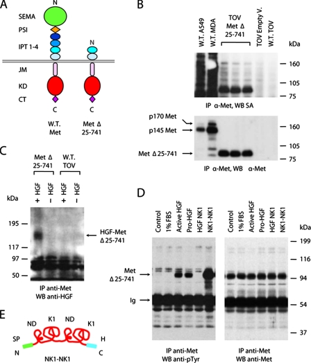
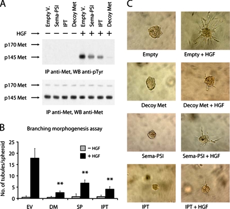
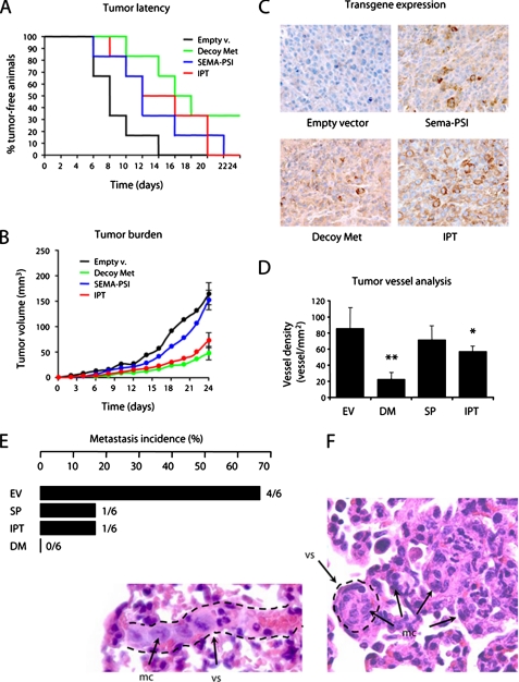
Hepatocyte growth factor (HGF) and its high affinity receptor, the tyrosine kinase Met, play a key role in embryo development and tumor invasion. Both HGF and Met are established targets for cancer therapy. However, the mechanism of their interaction is complex and remains elusive. HGF is secreted as a monomeric precursor (pro-HGF) that binds to but does not activate Met. Mature HGF is a alpha/beta heterodimer containing a high affinity Met-binding site in the alpha-chain (HGF-alpha) and a low affinity Met-binding site in the beta-chain (HGF-beta). The extracellular portion of Met contains a semaphorin (Sema) domain, a cysteine-rich hinge (plexin-semaphorin-integrin), and four immunoglobulin-like domains (immunoglobulin-like regions in plexins and transcription factors (IPT) 1-4). HGF-beta binds to Sema through a low affinity contact. The domain of Met responsible for high affinity binding to HGF-alpha has not been identified yet. Here we show that this long sought after binding site lies in the immunoglobulin-like region of Met and more precisely in IPT 3 and 4. We also show that IPT 3 and 4 are sufficient to transmit the signal for kinase activation to the cytoplasm, although the lack of Sema makes the receptor equally sensitive to mature HGF and pro-HGF. Finally, we provide evidence that soluble Met-derived proteins containing either the low affinity or high affinity HGF-binding site antagonize HGF-induced invasive growth both in vitro and in xenografts. These data suggest that the immunoglobulin-like region of Met cooperates with the Sema domain in binding to HGF and in controlling Met kinase activity. Although the IPT-HGF-alpha interaction provides binding strength, the Sema-HGF-beta contact confers selective sensitivity to the active form of the ligand.


Figures

FIGURE 1.
FIGURE 1.
Engineering and purification of Met and HGF subdomains. A, schematic representation of the engineered proteins used in this study. Left panel, engineered receptors. W.T. MET, wild-type Met; EXTRA, extracellular portion; INTRA, intracellular portion; SP, signal peptide; SEMA, semaphorin homology domain; PSI, plexin-semaphorin-integrin homology domain; IPT 1-4, immunoglobulin-plexin-transcription factor homology domains 1-4; TM, transmembrane domain; JM, juxta-membrane domain; KD, kinase domain; CT, C-terminal tail; E, FLAG or Myc epitope; H, polyhistidine tag. The red triangle indicates the proteolytic cleavage site between the α- and β-chain. Right panel, engineered ligands. W.T. HGF, wild-type HGF; ND, N-domain; K1-4, kringle 1-4; PLD, protease-like domain; UNCL. HGF, uncleavable HGF. The asterisk indicates the R494Q amino acid substitution in the proteolytic site. B, Coomassie staining of affinity-purified receptors and ligands. Each protein group (Sema, Sema-PSI, Decoy Met; PSI-IPT, IPT; HGF-α, Uncleavable HGF, HGF; HGF NK1, HGF-β) has been resolved by SDS-PAGE in nonreducing conditions. MW, molecular mass marker.
FIGURE 2.
FIGURE 2.
ELISA analysis of HGF-Met interactions. A, binding of Met subdomains to active HGF. Engineered receptors were immobilized in solid phase and exposed to increasing concentrations of active HGF in liquid phase. Binding was revealed using anti-HGF antibodies. Nonspecific binding was measured by using bovine serum albumin instead of purified receptors in solid phase. B-D, binding of Decoy Met, Sema-PSI, and IPT to different forms of HGF. Engineered receptors were immobilized in solid phase and exposed to increasing concentrations of Myc-tagged active HGF, pro-HGF, HGF-α, or HGF NK1 in liquid phase. Binding was revealed using anti-Myc antibodies. Nonspecific binding was measured by using Myc-tagged angiostatin (AS) in liquid phase.
FIGURE 3.
FIGURE 3.
IPT domains 3 and 4 are sufficient to binding to HGF-α at high affinity. A, schematic representation of deleted IPT variants. Color code and legend as in Fig. 1A. B, ELISA analysis of interactions between IPT variants and HGF-α. Engineered IPTs were immobilized in solid phase and exposed to increasing concentrations of HGF-α in liquid phase. Binding was revealed using biotinylated anti-HGF antibodies.
FIGURE 4.
FIGURE 4.
IPT domains 3 and 4 are sufficient for binding to HGF in living cells. A, schematic representation of the deleted MetΔ25-741 receptor. The color code and legend are as in Fig. 1A. B, surface biotinylation analysis. Cellular proteins were immunoprecipitated (IP) using antibodies directed against the C-terminal portion of Met and analyzed by Western blotting (WB) using horseradish peroxidase-conjugated streptavidin (SA). The same blots were reprobed with anti-Met antibodies. W.T., wild type; A549, A549 human lung carcinoma cells; MDA, MDA-MB-435 human melanoma cells; TOV, TOV-112D human ovary carcinoma cells; Empty V., empty vector. The p170 band corresponds to unprocessed Met; p145 is the mature form of the receptor. C, chemical cross-linking analysis. TOV-112D cells expressing MetΔ25-741 (Met Δ25-741) and wild-type TOV-112D cells (W.T. TOV) were incubated with HGF and then subjected to chemical cross-linking. The cell lysates were immunoprecipitated using anti-Met antibodies and analyzed by Western blotting using anti-HGF antibodies. The arrow indicates HGF-MetΔ25-741 complexes. D, Met phosphorylation analysis. TOV-112D cells expressing MetΔ25-741 were stimulated with 1% FBS as a negative control and with equal amounts of HGF, pro-HGF, HGF NK1, or NK1-NK1. Receptor phosphorylation was determined by immunoprecipitation with anti-Met antibodies and Western blotting with anti-phosphotyrosine (anti-pTyr) antibodies. The same blots were reprobed using anti-Met antibodies. The arrows indicate bands corresponding to MetΔ25-741 or immunoglobulins (Ig). E, schematic representation of NK1-NK1. From N to C termini: SP, signal peptide; ND, N-domain; K1, kringle 1; H, polyhistidine tag.
FIGURE 5.
FIGURE 5.
Soluble IPT inhibits HGF-induced invasive growth in vitro. A, lentiviral vector-transduced MDA-MB-435 cells were stimulated with recombinant HGF, and Met phosphorylation was determined by immunoblotting using anti-phosphotyrosine antibodies (upper panel). The same blot was reprobed using anti-Met antibodies (lower panel). Empty V., empty vector. B, branching morphogenesis assay. Preformed spheroids of lentiviral vector-transduced MDA-MB-435 cells were embedded in collagen and then stimulated with recombinant HGF to form branched tubules. Collagen invasion was quantified by scoring the mean number of tubules sprouting from each spheroid. EV, empty vector; DM, Decoy Met; SP, Sema-PSI. C, representative images from the experiment described in B. Magnification, 200×. IP, immunoprecipitation; WB, Western blotting.
FIGURE 6.
FIGURE 6.
Soluble IPT displays anti-tumor and anti-metastatic activity in mice. CD-1 nu-/- mice were injected subcutaneously with lentiviral vector-transduced MDA-MB-435 cells, and tumor growth was monitored over time. A, Kaplan-Meier-like plots of tumor latency (x axis, time in days; y axis, percent of tumor free-animals). Empty v., empty vector. B, mean tumor volume over time. C, immunohistochemical analysis of tumor sections using anti-FLAG antibodies. Magnification, 400×. D, tumor vessel analysis. Tumor sections were stained with anti-von Willebrand factor antibodies. The number of vessels per square mm of tumor section was determined by microscopy. EV, empty vector; DM, Decoy Met; SP, Sema-PSI. E, metastasis incidence analysis. Upon autopsy, serial lung sections were analyzed by microscopy to determine the presence of micrometastases. Metastasis incidence, i.e. the number of mice with metastasis over the total, is indicated in both percentage (bars) and fraction (at the ends of the bars). F, representative images of micrometastases from the empty vector group. Lung sections were stained with hematoxylin and eosin. The dotted lines identify the walls of blood vessels (vs). Metastatic cells (mc) can be found inside vessels as an embolus or in the parenchyma. Magnification, 400×.

References

    1. Bottaro, D. P., Rubin, J. S., Faletto, D. L., Chan, A. M., Kmiecik, T. E., Vande Woude, G. F., and Aaronson, S. A. (1991) Science 251 802-804 - PubMed
    1. Naldini, L., Vigna, E., Narsimhan, R. P., Gaudino, G., Zarnegar, R., Michalopoulos, G. K., and Comoglio, P. M. (1991) Oncogene 6 501-504 - PubMed
    1. Tempest, P. R., Stratton, M. R., and Cooper, C. S. (1988) Br. J. Cancer 58 3-7 - PMC - PubMed
    1. Giordano, S., Ponzetto, C., Di Renzo, M. F., Cooper, C. S., and Comoglio, P. M. (1989) Nature 339 155-156 - PubMed
    1. Komada, M., Hatsuzawa, K., Shibamoto, S., Ito, F., Nakayama, K., and Kitamura, N. (1993) FEBS Lett. 328 25-29 - PubMed

Publication types