

Identification and analysis of conserved sequence motifs in cytochrome P450 family 2. Functional and structural role of a motif 187RFDYKD192 in CYP2B enzymes
- PMID: 18495666
- PMCID: PMC2490781
- DOI: 10.1074/jbc.M708582200
Identification and analysis of conserved sequence motifs in cytochrome P450 family 2. Functional and structural role of a motif 187RFDYKD192 in CYP2B enzymes
Abstract
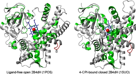
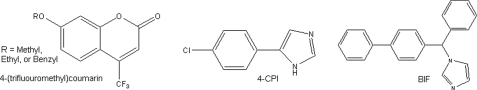
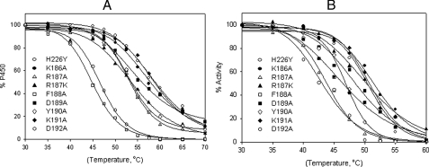
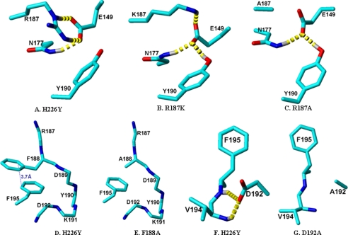
Using a multiple alignment of 175 cytochrome P450 (CYP) family 2 sequences, 20 conserved sequence motifs (CSMs) were identified with the program PCPMer. Functional importance of the CSM in CYP2B enzymes was assessed from available data on site-directed mutants and genetic variants. These analyses suggested an important role of the CSM 8, which corresponds to(187)RFDYKD(192) in CYP2B4. Further analysis showed that residues 187, 188, 190, and 192 have a very high rank order of conservation compared with 189 and 191. Therefore, eight mutants (R187A, R187K, F188A, D189A, Y190A, K191A, D192A, and a negative control K186A) were made in an N-terminal truncated and modified form of CYP2B4 with an internal mutation, which is termed 2B4dH/H226Y. Function was examined with the substrates 7-methoxy-4-(trifluoromethyl)coumarin (7-MFC), 7-ethoxy-4-(trifluoromethyl)coumarin (7-EFC), 7-benzyloxy-4-(trifluoromethyl)coumarin (7-BFC), and testosterone and with the inhibitors 4-(4-chlorophenyl)imidazole (4-CPI) and bifonazole (BIF). Compared with the template and K186A, the mutants R187A, R187K, F188A, Y190A, and D192A showed > or =2-fold altered substrate specificity, k(cat), K(m), and/or k(cat)/K(m) for 7-MFC and 7-EFC and 3- to 6-fold decreases in differential inhibition (IC(50,BIF)/IC(50,4-CPI)). Subsequently, these mutants displayed 5-12 degrees C decreases in thermal stability (T(m)) and 2-8 degrees C decreases in catalytic tolerance to temperature (T(50)) compared with the template and K186A. Furthermore, when R187A and D192A were introduced in CYP2B1dH, the P450 expression and thermal stability were decreased. In addition, R187A showed increased activity with 7-EFC and decreased IC(50,BIF)/IC(50,4-CPI) compared with 2B1dH. Analysis of long range residue-residue interactions in the CYP2B4 crystal structures indicated strong hydrogen bonds involving Glu(149)-Asn(177)-Arg(187)-Tyr(190) and Asp(192)-Val(194), which were significantly-reduced/abolished by the Arg(187)-->Ala and Asp(192)-->Alasubstitutions, respectively.
Figures
Similar articles
-
Investigation of the role of cytochrome P450 2B4 active site residues in substrate metabolism based on crystal structures of the ligand-bound enzyme.Arch Biochem Biophys. 2006 Nov 1;455(1):61-7. doi: 10.1016/j.abb.2006.08.024. Epub 2006 Sep 25. Arch Biochem Biophys. 2006. PMID: 17027909 Free PMC article.
-
Coumarin Derivatives as Substrate Probes of Mammalian Cytochromes P450 2B4 and 2B6: Assessing the Importance of 7-Alkoxy Chain Length, Halogen Substitution, and Non-Active Site Mutations.Biochemistry. 2016 Apr 5;55(13):1997-2007. doi: 10.1021/acs.biochem.5b01330. Epub 2016 Mar 24. Biochemistry. 2016. PMID: 26982502 Free PMC article.
-
Investigation by site-directed mutagenesis of the role of cytochrome P450 2B4 non-active-site residues in protein-ligand interactions based on crystal structures of the ligand-bound enzyme.FEBS J. 2012 May;279(9):1607-20. doi: 10.1111/j.1742-4658.2011.08411.x. Epub 2011 Nov 25. FEBS J. 2012. PMID: 22051155 Free PMC article.
-
Structure-function analysis of cytochromes P450 2B.Biochim Biophys Acta. 2007 Mar;1770(3):402-12. doi: 10.1016/j.bbagen.2006.07.006. Epub 2006 Jul 22. Biochim Biophys Acta. 2007. PMID: 16935426 Review.
-
Plasticity of CYP2B enzymes: structural and solution biophysical methods.Curr Drug Metab. 2012 Feb;13(2):167-76. doi: 10.2174/138920012798918417. Curr Drug Metab. 2012. PMID: 22208531 Free PMC article. Review.
Cited by
-
Decreased susceptibility of the cytochrome P450 2B6 variant K262R to inhibition by several clinically important drugs.Drug Metab Dispos. 2009 Mar;37(3):644-50. doi: 10.1124/dmd.108.023655. Epub 2008 Dec 12. Drug Metab Dispos. 2009. PMID: 19074527 Free PMC article.
-
Engineering proteins with enhanced mechanical stability by force-specific sequence motifs.Proteins. 2012 May;80(5):1308-15. doi: 10.1002/prot.24027. Epub 2012 Feb 10. Proteins. 2012. PMID: 22274941 Free PMC article.
-
Potent mechanism-based inactivation of cytochrome P450 2B4 by 9-ethynylphenanthrene: implications for allosteric modulation of cytochrome P450 catalysis.Biochemistry. 2013 Jan 15;52(2):355-64. doi: 10.1021/bi301567z. Epub 2013 Jan 4. Biochemistry. 2013. PMID: 23276288 Free PMC article.
-
Interactions between CYP2E1 and CYP2B4: effects on affinity for NADPH-cytochrome P450 reductase and substrate metabolism.Drug Metab Dispos. 2013 Jan;41(1):101-10. doi: 10.1124/dmd.112.046094. Epub 2012 Oct 5. Drug Metab Dispos. 2013. PMID: 23043184 Free PMC article.
-
Substrate tunnels in enzymes: structure-function relationships and computational methodology.Proteins. 2015 Apr;83(4):599-611. doi: 10.1002/prot.24772. Epub 2015 Feb 28. Proteins. 2015. PMID: 25663659 Free PMC article. Review.
References
Publication types
MeSH terms
Substances
Grants and funding
LinkOut - more resources
Full Text Sources
Miscellaneous
