. 2008 Aug;82(15):7298-305.
doi: 10.1128/JVI.00772-08. Epub 2008 May 21.

Dimerization of the papillomavirus E2 protein is required for efficient mitotic chromosome association and Brd4 binding

Affiliations

Dimerization of the papillomavirus E2 protein is required for efficient mitotic chromosome association and Brd4 binding

Juan Cardenas-Mora et al. J Virol. 2008 Aug.

Abstract

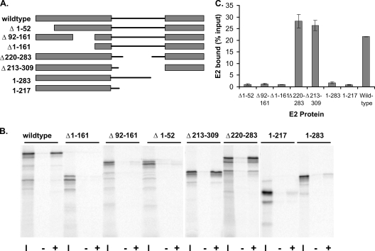
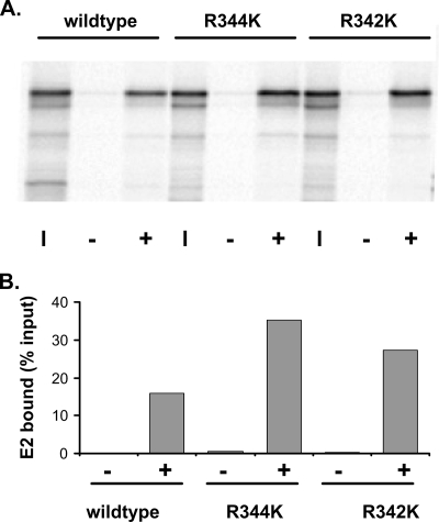
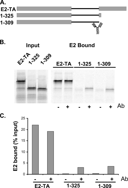
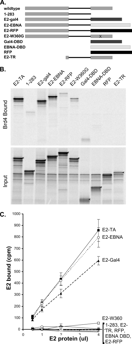
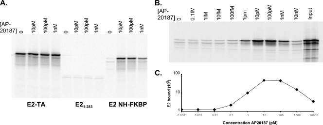
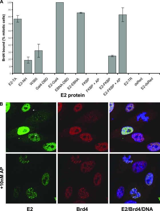
The E2 proteins of several papillomaviruses link the viral genome to mitotic chromosomes to ensure retention and the efficient partitioning of genomes into daughter cells following cell division. Bovine papillomavirus type 1 E2 binds to chromosomes in a complex with Brd4, a cellular bromodomain protein. Interaction with Brd4 is also important for E2-mediated transcriptional regulation. The transactivation domain of E2 is crucial for interaction with the Brd4 protein; proteins lacking or mutated in this domain do not interact with Brd4. However, we found that the C-terminal DNA binding/dimerization domain of E2 is also required for efficient binding to Brd4. Mutations that eliminated the DNA binding function of E2 had no effect on the ability of E2 to interact with Brd4, but an E2 protein with a mutation that disrupted C-terminal dimerization bound Brd4 with greatly reduced efficiency. Furthermore, E2 proteins in which the C-terminal domains were replaced with the dimeric DNA binding domain of EBNA-1 or Gal4 bound efficiently to the Brd4 protein, but the replacement of the E2 C-terminal domain with a monomeric red fluorescent protein did not rescue efficient Brd4 binding. Thus, E2 bound to Brd4 most efficiently as a dimer. To prove this finding further, the E2 DNA binding domain was replaced with an FKBP12-derived domain in which dimerization was regulated by a bivalent ligand. This fusion protein bound Brd4 efficiently only in the presence of the ligand, confirming that a dimer of E2 was required. Correspondingly, E2 proteins that could dimerize were able to bind to mitotic chromosomes much more efficiently than monomeric E2 polypeptides.


Figures

FIG. 1.
FIG. 1.
Efficient interaction of E2 and Brd4 requires both the transactivation and DNA binding/dimerization domains. (A) Diagram of truncated and deletion mutant E2 proteins used in the Brd4 binding assay. (B) In vitro-translated E2 proteins were tested for their abilities to bind Brd4. Aliquots (10 μl) of reticulocyte lysate (adjusted for the concentration of E2) were assayed for binding to 5 μl of Brd4 protein extract prebound to anti-FLAG immunobeads. Bound proteins were eluted and analyzed by SDS-PAGE. The input lanes (I) contain a 1/4 volume of lysate added to the binding reactions. The lanes labeled − show E2 bound in the absence of Brd4, and the lanes labeled + show E2 bound in the presence of Brd4 protein. (C) Percentage of E2 bound for each E2 protein, as obtained from two independent assays.
FIG. 2.
FIG. 2.
The E2 DNA binding function is not required for Brd4 binding. (A) E2 proteins that are unable to bind specifically to DNA were tested in the Brd4 binding assay, as described in the legend to Fig. 1. The input lanes (I) contain lysate added to the reaction mixture at a ratio of 1/4. The lanes labeled − show E2 bound in the absence of Brd4, and the lanes labeled + show E2 bound in the presence of Brd4 protein. R344K and R342K, E2 mutant proteins with the R344K and R342K substitutions, respectively. (B) The amount of E2 bound was quantitated using a Molecular Dynamics Typhoon imager.
FIG. 3.
FIG. 3.
Antibody-mediated dimerization enhances the ability of E2 to bind to Brd4. (A) Diagram of truncated E2 proteins used in the Brd4 binding assay. The cartoon of an antibody molecule indicates the position of the B202 epitope. E2-TA, wild-type E2 protein. (B) In vitro-translated E2 proteins were tested for their abilities to bind Brd4 in the absence or presence of the B202 antibody. The panel on the left shows results for a 1/4 volume of lysate added to the binding reactions. The panel on the right shows the bound proteins in the absence (−) and presence (+) of the B202 antibody (Ab). (C) The amount of E2 bound was quantitated using a Molecular Dynamics Typhoon imager.
FIG. 4.
FIG. 4.
The efficient interaction of E2 and Brd4 requires E2 dimerization. (A) Diagram of truncated, deletion mutant, and fusion E2 proteins used in the Brd4 binding assay. DBD, DNA binding domain; E2-TR, E2 repressor protein. (B) In vitro-translated E2 proteins were tested for their abilities to bind Brd4. Aliquots (4 μl) of reticulocyte lysate (adjusted for the concentration of E2) were assayed for binding to 5 μl of Brd4 protein extract prebound to anti-FLAG immunobeads. Bound proteins were eluted and analyzed by SDS-PAGE. The input lanes contain a 1/4 volume of lysate added to the binding reactions. E2-TA, wild-type E2 protein. (C) The efficiency of E2-Brd4 binding was quantitated by adding increasing volumes of reticulocyte lysate (adjusted for the concentration of E2) to Brd4 beads. The amount bound was determined directly by scintillation counting.
FIG. 5.
FIG. 5.
(A) In vitro-translated E2 proteins were tested for their abilities to bind Brd4 in the presence of increasing amounts of AP20187. Aliquots (4 μl) of reticulocyte lysate (adjusted for the concentration of E2) were assayed for binding to 5 μl of Brd4 protein extract prebound to anti-FLAG immunobeads. Bound proteins were eluted and analyzed by SDS-PAGE. E2-TA, wild-type E2 protein. (B) The optimal amount of AP20187 required to enhance E2-FKBP E2 binding to Brd4 was determined as described in the legend to panel A. Bound E2-FKBP was quantitated using a Molecular Dynamics Typhoon imager. Levels of bound E2 are expressed as percentages relative to the input.
FIG. 6.
FIG. 6.
(A) FLAG-tagged E2 proteins were expressed in CV-1 cell lines, and the level of each protein was determined by immunoblot analysis using an anti-FLAG M2 antibody. E2-TA, wild-type E2 protein; E2-N, E2 N terminus; DBD, DNA binding domain; E2-TR, E2 repressor protein. (B) E2 W360G is defective in dimerization. Nuclear extracts from CV-1 cells expressing FLAG-tagged wild-type BPV-1 E2 (WT) and W360G proteins were fractionated by Superdex 200 10/300 GL column chromatography. The fractions were analyzed by immunoblotting using anti-FLAG M2 antibody. Gel filtration standards (Bio-Rad) were run in parallel to independently assess the sizes of the complexes and are indicated in kilodaltons. (C) FLAG-tagged E2-FKBP fusion proteins were expressed in CV-1 cell lines, and the level of each protein in the presence (+) or absence (−) of 10 nM AP20187 was determined by immunoblot analysis using an anti-FLAG M2 antibody.
FIG. 7.
FIG. 7.
(A) The E2 proteins indicated were expressed in CV-1 cells, and the localization of each E2 protein in a complex with Brd4 was detected by indirect immunofluorescence. The percentage of mitotic E2-expressing cells that had recruited Brd4 in speckles to the mitotic chromosomes is shown, along with the standard deviation of results derived from several experiments. The bars labeled +AP show results for cells treated with 10 nM AP20187. E2-TA, wild-type E2 protein; DBD, DNA binding domain; E2-TR, E2 repressor protein. (B) Representative indirect immunofluorescence images of E2-FKBP proteins (green), including proteins in complexes with Brd4 (red), on mitotic chromosomes in the presence or absence of 10 nM AP20187. Cellular chromosomes are stained with DAPI (blue). Mitotic cells are indicated by arrows.

References

    1. Abbate, E. A., C. Voitenleitner, and M. R. Botchan. 2006. Structure of the papillomavirus DNA-tethering complex E2:Brd4 and a peptide that ablates HPV chromosomal association. Mol. Cell 24877-889. - PubMed
    1. Androphy, E. J., D. R. Lowy, and J. T. Schiller. 1987. Bovine papillomavirus E2 trans-activating gene product binds to specific sites in papillomavirus DNA. Nature 32570-73. - PubMed
    1. Antson, A. A., J. E. Burns, O. V. Moroz, D. J. Scott, C. M. Sanders, I. B. Bronstein, G. G. Dodson, K. S. Wilson, and N. J. Maitland. 2000. Structure of the intact transactivation domain of the human papillomavirus E2 protein. Nature 403805-809. - PubMed
    1. Bastien, N., and A. A. McBride. 2000. Interaction of the papillomavirus E2 with mitotic chromosomes. Virology 270124-134. - PubMed
    1. Baxter, M. K., M. G. McPhillips, K. Ozato, and A. A. McBride. 2005. The mitotic chromosome binding activity of the papillomavirus E2 protein correlates with interaction with the cellular chromosomal protein, Brd4. J. Virol. 794806-4818. - PMC - PubMed

Publication types

LinkOut - more resources