

Transmission of human immunodeficiency virus type 1 from a patient who developed AIDS to an elite suppressor
- PMID: 18495769
- PMCID: PMC2493308
- DOI: 10.1128/JVI.00800-08
Transmission of human immunodeficiency virus type 1 from a patient who developed AIDS to an elite suppressor
Abstract
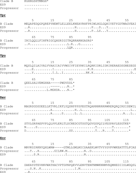
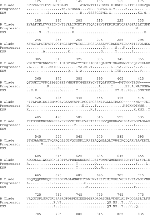
Elite suppressors (ES) are untreated human immunodeficiency virus type 1 (HIV-1)-infected patients who maintain viral loads of <50 copies/ml. The mechanisms involved in this control of viral replication remain unclear. Prior studies suggested that these patients, as well as long-term nonprogressors, are infected with defective HIV-1 variants. Other reports have shown that the HLA-B*27 and -B*57 alleles are overrepresented in these patients, suggesting that host factors play a role in the control of viral replication. In order to distinguish between these hypotheses, we studied differences in viral isolates and immune responses of an HIV-1 transmission pair. While both patients are HLA-B*57 positive, the transmitter progressed to AIDS, whereas the recipient, who is also HLA-B*27 positive, is an ES. Isolates from both patients were replication competent and contained the T242N escape mutation in Gag, which is known to decrease viral fitness. While the acquisition of compensatory mutations occurred in isolates from the progressor, a superior HIV-specific CD8(+) T-cell response in the ES appears to have prevented viral replication and thus the evolution toward a more fit variant. In addition, CD8(+) T cells in the ES have selected for a rare mutation in an immunodominant HLA-B*27-restricted Gag epitope, which also has a negative impact on fitness. The results strongly suggest that through direct and indirect mechanisms, CD8(+) T cells in some ES control HIV-1 isolates are capable of causing profound immunosuppression.
Figures


References
-
- Addo, M. M., X. G. Yu, A. Rathod, D. Cohen, R. L. Eldridge, D. Strick, M. N. Johnston, C. Corcoran, A. G. Wurcel, C. A. Fitzpatrick, M. E. Feeney, W. R. Rodriguez, N. Basgoz, R. Draenert, D. R. Stone, C. Brander, P. J. R. Goulder, E. S. Rosenberg, M. Altfeld, and B. D. Walker. 2003. Comprehensive epitope analysis of human immunodeficiency virus type 1 (HIV-1)-specific T-cell responses directed against the entire expressed HIV-1 genome demonstrate broadly directed responses, but no correlation to viral load. J. Virol. 772081-2092. - PMC - PubMed
-
- Alexander, L., E. Weiskopf, T. C. Greenough, N. C. Gaddis, M. R. Auerbach, M. H. Malim, S. J. O'Brien, B. D. Walker, J. L. Sullivan, and R. C. Desrosiers. 2000. Unusual polymorphisms in human immunodeficiency virus type 1 associated with nonprogressive infection. J. Virol. 744361-4376. - PMC - PubMed
-
- Almeida, J. R., D. A. Price, L. Papagno, Z. A. Arkoub, D. Sauce, E. Bornstein, T. E. Asher, A. Samri, A. Schnuriger, I. Theodorou, D. Costagliola, C. Rouzioux, H. Agut, A. G. Marcelin, D. Douek, B. Autran, and V. Appay. 2007. Superior control of HIV-1 replication by CD8+ T cells is reflected by their avidity, polyfunctionality, and clonal turnover. J. Exp. Med. 2042473-2485. - PMC - PubMed
-
- Alter, G., M. P. Martin, N. Teigen, W. H. Carr, T. J. Suscovich, A. Schneidewind, H. Streeck, M. Waring, A. Meier, C. Brander, J. D. Lifson, T. M. Allen, M. Carrington, and M. Altfeld. 2007. Differential natural killer cell-mediated inhibition of HIV-1 replication based on distinct KIR/HLA subtypes. J. Exp. Med. 2043027-3036. - PMC - PubMed
-
- Bailey, J. R., K. G. Lassen, H. C. Yang, T. C. Quinn, S. C. Ray, J. N. Blankson, and R. F. Siliciano. 2006. Neutralizing antibodies do not mediate suppression of human immunodeficiency virus type 1 in elite suppressors or selection of plasma virus variants in patients on highly active antiretroviral therapy. J. Virol. 804758-4770. - PMC - PubMed
Publication types
MeSH terms
Substances
Associated data
- Actions
- Actions
Grants and funding
LinkOut - more resources
Full Text Sources
Other Literature Sources
Medical
Research Materials
