. 2008 Jul;295(1):F215-25.
doi: 10.1152/ajprenal.00548.2007. Epub 2008 May 21.

Protein kinase B/Akt activity is involved in renal TGF-beta1-driven epithelial-mesenchymal transition in vitro and in vivo

Affiliations

Protein kinase B/Akt activity is involved in renal TGF-beta1-driven epithelial-mesenchymal transition in vitro and in vivo

Jayesh J Kattla et al. Am J Physiol Renal Physiol. 2008 Jul.

Abstract

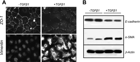
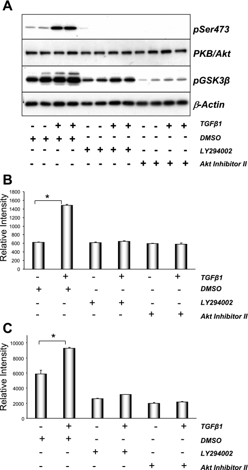
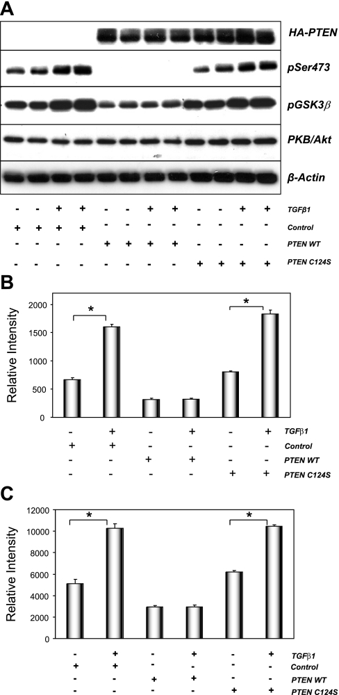
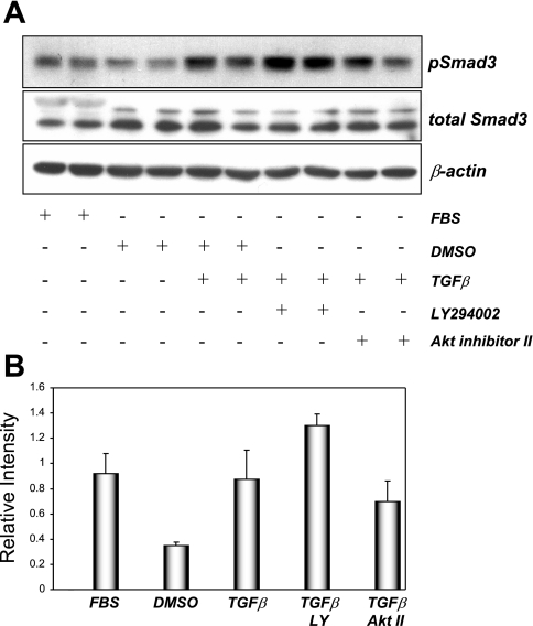
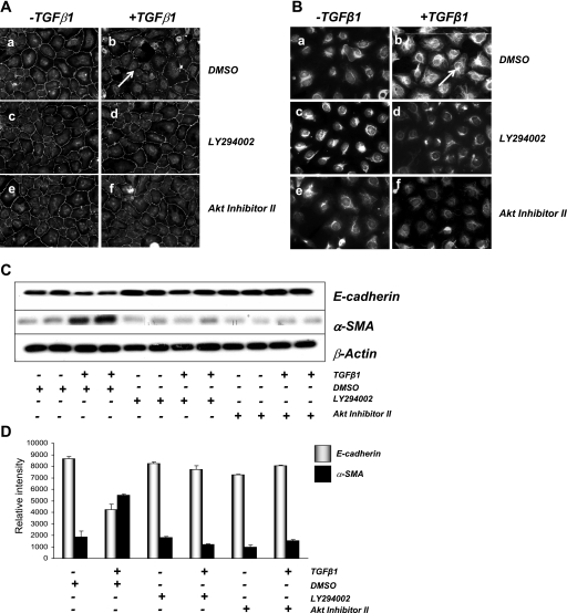
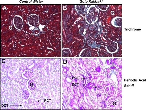
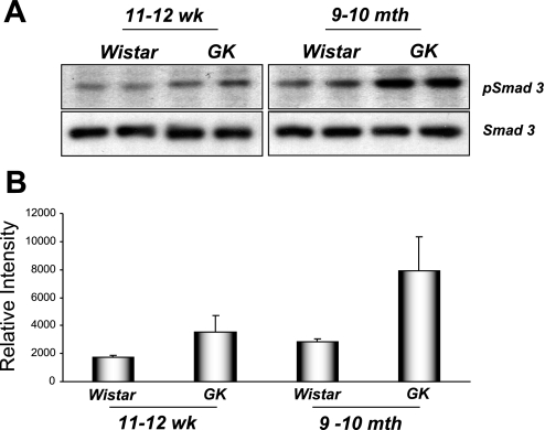
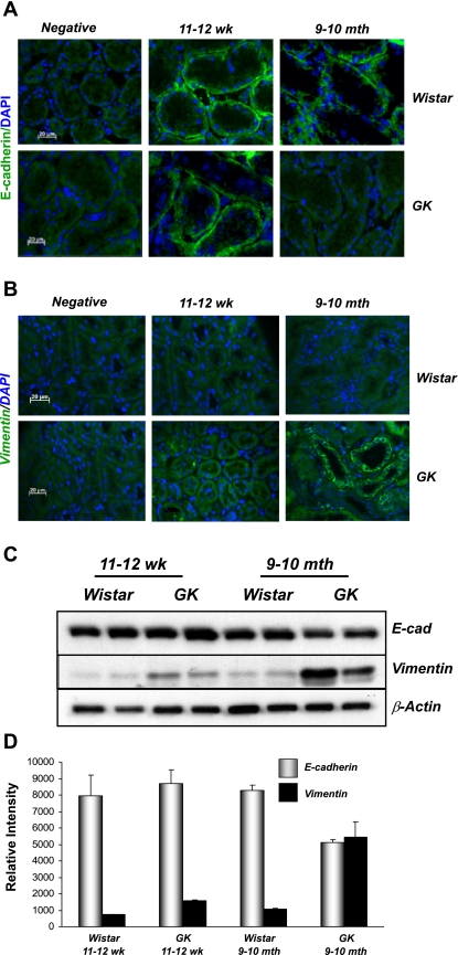
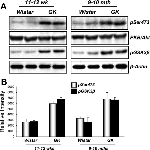
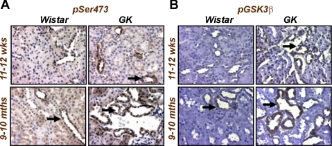
The molecular pathogenesis of diabetic nephropathy (DN), the leading cause of end-stage renal disease worldwide, is complex and not fully understood. Transforming growth factor-beta (TGF-beta1) plays a critical role in many fibrotic disorders, including DN. In this study, we report protein kinase B (PKB/Akt) activation as a downstream event contributing to the pathophysiology of DN. We investigated the potential of PKB/Akt to mediate the profibrotic bioactions of TGF-beta1 in kidney. Treatment of normal rat kidney epithelial cells (NRK52E) with TGF-beta1 resulted in activation of phosphatidylinositol 3-kinase (PI3K) and PKB/Akt as evidenced by increased Ser473 phosphorylation and GSK-3beta phosphorylation. TGF-beta1 also stimulated increased Smad3 phosphorylation in these cells, a response that was insensitive to inhibition of PI3K or PKB/Akt. NRK52E cells displayed a loss of zona occludins 1 and E-cadherin and a gain in vimentin and alpha-smooth muscle actin expression, consistent with the fibrotic actions of TGF-beta1. These effects were blocked with inhibitors of PI3K and PKB/Akt. Furthermore, overexpression of PTEN, the lipid phosphatase regulator of PKB/Akt activation, inhibited TGF-beta1-induced PKB/Akt activation. Interestingly, in the Goto-Kakizaki rat model of type 2 diabetes, we also detected increased phosphorylation of PKB/Akt and its downstream target, GSK-3beta, in the tubules, relative to that in control Wistar rats. Elevated Smad3 phosphorylation was also detected in kidney extracts from Goto-Kakizaki rats with chronic diabetes. Together, these data suggest that TGF-beta1-mediated PKB/Akt activation may be important in renal fibrosis during diabetic nephropathy.


Figures

Fig. 1.
Fig. 1.
Transforming growth factor-β1 (TGF-β1) induces tubular epithelial-mesenchymal transition (EMT) in NRK52E normal rat kidney epithelial cells. A: NRK52E proximal tubule epithelial cells were grown on glass coverslips in the presence of vehicle (left) or 10 ng/ml TGF-β1 (right) for 6 days. Cells were fixed using 4% (wt/vol) paraformaldehyde and incubated with primary antibodies against zona occludins 1 (ZO-1) to visualize cell junctions or vimentin to visualize cytoskeleton as described in materials and methods. Immunoreactivity was visualized using TRITC-labeled secondary antibodies, and nuclei were visualized using 4,6-diamidino-2-phenylindole (DAPI)/Hoechst 33258 at ×200 magnification. B: Western blot analysis of extracts (25 μg protein/lane) from NRK52E cells incubated in the presence or absence of 10 ng/ml TGF-β1. Lysates were probed for E-cadherin, α-smooth muscle actin (α-SMA), or β-actin overnight.
Fig. 2.
Fig. 2.
TGF-β1 induced phosphatidylinositol 3-kinase (PI3K)→PKB/Akt signaling in NRK52E cells. A: serum-starved NRK52E cells were pretreated with DMSO, 20 μM LY-294002, or 40 μM Akt inhibitor II for 30 min. Cells were then treated with vehicle or 10 ng/ml TGF-β1 for 60 min. Protein lysate (25 μg) was probed using antibodies against activated PKB/Akt (pSer473), total Akt protein (PKB/Akt), phosphorylated GSK-3β (pSer9), or β-actin. Starved NRK52E cells were processed and proteins extracts quantified for pAkt (B) and pGSK-3β Ser9 (C) using Meso Scale Discovery (MSD) electrochemiluminescence technology. Values for pAkt and pGSK-3β were normalized to their respective total protein levels. Data are means ± SE; n = 4. *P < 0.0005; paired t-test.
Fig. 3.
Fig. 3.
Wild-type but not mutant PTEN expression blocks TGF-β-mediated PKB/Akt activation in NRK52E cells. A: NRK52E cells were transfected with hemagglutinin (HA)-tagged pCGN empty vector (control), pCGN-PTEN wild type (PTEN WT), or pCGN-PTEN C124S phosphatase inactive mutant (PTEN C124S). Transfected cells were then cultured in the presence of vehicle or 10 ng/ml TGF-β1 for 24 h. Protein extracts (25 μg) were probed for HA-PTEN or HA-PTEN C124S, phosphorylated PKB/Akt and GSK-3β, total PKB/Akt, and β-actin as described. NRK52E cells were processed and proteins extracts quantified for pSer473 (B) and pGSK-3β Ser9 (C) using MSD electrochemiluminescence technology. Values represent the ratio of phosphoprotein to total protein (see materials and methods). Data are means ± SE; n = 4. *P < 0.0005; paired t-test.
Fig. 4.
Fig. 4.
TGF-β1-stimulated Smad3 phosphorylation in NRK52E cells is insensitive to PI3K inhibition. Serum-starved NRK52E cells were treated with DMSO, 20 μM LY-294002, or 20 μM Akt inhibitor II for 30 min before treatment with vehicle or 10 ng/ml TGF-β1 for 60 min at 37°C. Nonstarved cells (FBS) were also included in this experiment. Protein extracts (25 μg) were probed using antibodies raised against pSmad3, total Smad3, or β-actin as loading control. Values are representative of 3 experiments carried out in duplicate. Band intensities were calculated using Scion Image software. Data are means ± SD.
Fig. 5.
Fig. 5.
TGF-β1-mediated phosphorylation of PKB/Akt and GSK-3β is important for EMT in rat kidney cells. NRK52E cells were treated with DMSO, LY-294002 or Akt inhibitor II for 30 min before treatment with 10 ng/ml TGF-β1 for 72 h. Cells were stained for epithelial marker ZO-1 (A) and mesenchymal marker vimentin (B). Cells were fixed using 4% (wt/vol) paraformaldehyde, and immunoreactivity was visualized using TRITC-labeled secondary antibodies at ×200 magnification using fluorescence microscopy. C: NRK52E cells were exposed to DMSO, 20 μM LY-294002, and 40 μM Akt inhibitor II in the presence of vehicle or 10 ng/ml TGF-β1 for 72 h. Protein extracts (25 μg) were probed using antibodies against E-cadherin, α-SMA, or β-actin. D: immunoblots were analyzed using densitometry (Scion Image software). The ratios of E-cadherin and α-SMA to β-actin were calculated. This experiment was carried out in triplicate with similar results. Data are means ± SD.
Fig. 6.
Fig. 6.
Diabetic Goto-Kakizaki (GK) rats display mild to moderate tubulointerstitial fibrosis at 9–10 mo of age. Kidney sections (5 μm) from nondiabetic control Wistar (A and C) or diabetic GK rats (B and D) (n = 4) were stained with Masson's Trichrome for collagen (A and B) or periodic acid-Schiff for extracellular matrix proteins (C and D) as described in materials and methods and examined at ×400 magnification. The locations of proximal convoluted tubule (PCT) and distal convoluted tubule (DCT) are indicated. Arrows pinpoint moderate tubulointerstitial fibrosis with collagen deposition (B) and thickening of the tubular basement membrane (D) in diabetic GK but not in control Wistar rats. Glomeruli are also marked (G).
Fig. 7.
Fig. 7.
Evidence of elevated TGF-β1 signaling in diabetic rata kidneys. Active TGF-β1 signaling was examined by immunoblotting for Smad3, a phosphorylated downstream target of TGF-β1. A: 25-μg protein extracts from 11- to 12-wk and 9- to 10-mo-old control and GK rat kidneys were probed for pSmad3 and total Smad3 using standard immunoblotting techniques. B: densitometry using Scion Image software was performed, and the intensity of pSmad3 bands were normalized to total Smad3 expression.
Fig. 8.
Fig. 8.
Evidence of EMT in GK diabetic rats. Fresh frozen sections (5 μm) from control Wistar or GK rats ages 11–12 wk or 9–10 mo were incubated with primary antibodies against E-cadherin (A) or vimentin (B) as described in materials and methods. Immunoreactivity (green) was visualized using FITC-labeled anti-mouse secondary antibodies, and nuclei were visualized using DAPI/Hoechst 33258 (blue). A control in which the primary antibody was replaced with blocking buffer was included in each experiment (negative). Sections were visualized at ×400 magnification. C and D: protein lysates from control Wistar or GK rats were probed for EMT markers. Protein extracts (25 μg) were probed using antibodies against E-cadherin, α-SMA, and β-actin (C). Band intensities were quantified using Scion Image software. The intensity ratios of E-cadherin and α-SMA to β-actin are shown (D). Data are means ± SD.
Fig. 9.
Fig. 9.
PKB/Akt pathway signaling is elevated in GK rats. A: protein extracts from control Wistar rats and GK rat kidneys were probed with antisera raised against pSer473 of PKB/Akt or pSer9 of GSK-3β. Protein loading was controlled using a polyclonal antibody raised against total PKB/Akt and β-actin, respectively. Data are representative of 4 rats from both control and GK groups at ages 11–12 wk and 9–10 mo. B: band intensities were calculated using Scion Image software. Data are means (SD).
Fig. 10.
Fig. 10.
Elevated PKB/Akt and GSK-3β localize to the kidney tubules of diabetic rats. Paraffin-embedded kidney sections (5 μm) from control Wistar and diabetic GK rats ages 11–12 wk and 9–10 mo were incubated with a rabbit polyclonal antibody against pSer473 of PKB/Akt (A) or pSer9 of GSK-3β (B) as described in materials and methods. Immunoreactivity was visualized using diaminobenzidine staining at ×400 magnification. Arrows indicate staining in the proximal and distal tubules.

References

    1. Bachelder RE, Yoon SO, Franci C, de Herreros AG, Mercurio AM. Glycogen synthase kinase-3 is an endogenous inhibitor of Snail transcription: implications for the epithelial-mesenchymal transition. J Cell Biol 168: 29–33, 2005. - PMC - PubMed
    1. Bakin AV, Rinehart C, Tomlinson AK, Arteaga CL. p38 mitogen-activated protein kinase is required for TGFβ-mediated fibroblastic transdifferentiation and cell migration. J Cell Sci 115: 3193–3206, 2002. - PubMed
    1. Bakin AV, Tomlinson AK, Bhowmick NA, Moses HL, Arteaga CL. Phosphatidylinositol 3-kinase function is required for transforming growth factor beta-mediated epithelial to mesenchymal transition and cell migration. J Biol Chem 275: 36803–36810, 2000. - PubMed
    1. Batlle E, Sancho E, Franci C, Dominguez D, Monfar M, Baulida J, Garcia De Herreros A. The transcription factor snail is a repressor of E-cadherin gene expression in epithelial tumour cells. Nat Cell Biol 2: 84–89, 2000. - PubMed
    1. Bhowmick NA, Ghiassi M, Bakin A, Aakre M, Lundquist CA, Engel ME, Arteaga CL, Moses HL. Transforming growth factor-beta1 mediates epithelial to mesenchymal transdifferentiation through a RhoA-dependent mechanism. Mol Biol Cell 12: 27–36, 2001. - PMC - PubMed

Publication types

MeSH terms