. 2008 Jul;295(1):C231-41.
doi: 10.1152/ajpcell.00175.2008. Epub 2008 May 21.

Rho-family GTPases modulate Ca(2+) -dependent ATP release from astrocytes

Affiliations

Rho-family GTPases modulate Ca(2+) -dependent ATP release from astrocytes

Andrew E Blum et al. Am J Physiol Cell Physiol. 2008 Jul.

Abstract

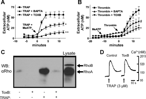
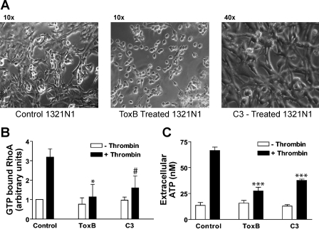
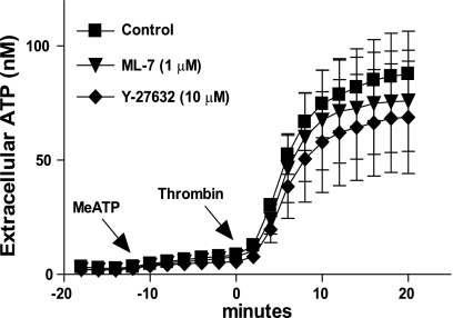
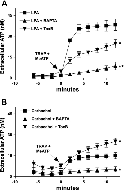
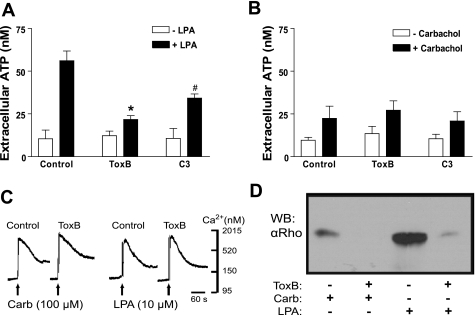
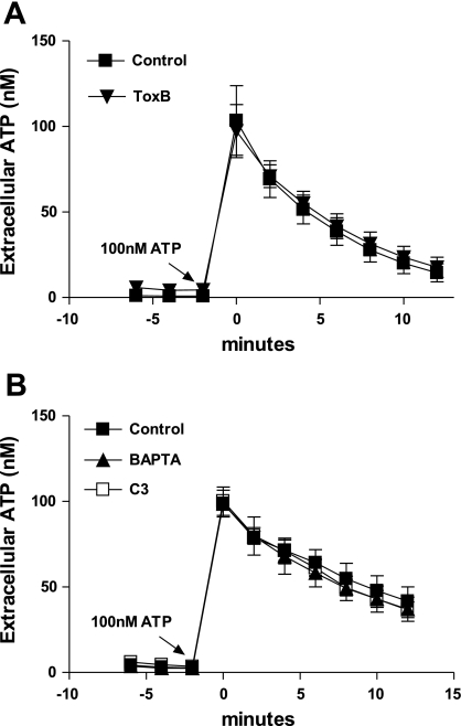
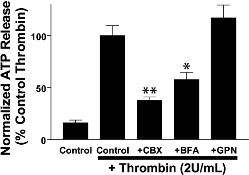
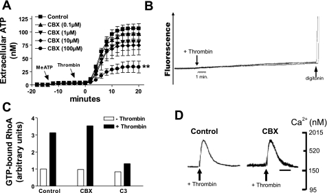
Previously, we reported that activation of G protein-coupled receptors (GPCR) in 1321N1 human astrocytoma cells elicits a rapid release of ATP that is partially dependent on a G(q)/phophospholipase C (PLC)/Ca(2+) mobilization signaling cascade. In this study we assessed the role of Rho-family GTPase signaling as an additional pathway for the regulation of ATP release in response to activation of protease-activated receptor-1 (PAR1), lysophosphatidic acid receptor (LPAR), and M3-muscarinic (M3R) GPCRs. Thrombin (or other PAR1 peptide agonists), LPA, and carbachol triggered quantitatively similar Ca(2+) mobilization responses, but only thrombin and LPA caused rapid accumulation of active GTP-bound Rho. The ability to elicit Rho activation correlated with the markedly higher efficacy of thrombin and LPA, relative to carbachol, as ATP secretagogues. Clostridium difficile toxin B and Clostridium botulinum C3 exoenzyme, which inhibit Rho-GTPases, attenuated the thrombin- and LPA-stimulated ATP release but did not decrease carbachol-stimulated release. Thus the ability of certain G(q)-coupled receptors to additionally stimulate Rho-GTPases acts to strongly potentiate a Ca(2+)-activated ATP release pathway. However, pharmacological inhibition of Rho kinase I/II or myosin light chain kinase did not attenuate ATP release. PAR1-induced ATP release was also reduced twofold by brefeldin treatment suggesting the possible mobilization of Golgi-derived, ATP-containing secretory vesicles. ATP release was also markedly repressed by the gap junction channel inhibitor carbenoxolone in the absence of any obvious thrombin-induced change in membrane permeability indicative of hemichannel gating.


Figures

Fig. 1.
Fig. 1.
Protease-activated receptor (PAR1)-mediated ATP release is sensitive to BAPTA and Clostridium difficile toxin B (ToxB). A: changes in stimulated extracellular [ATP] were recorded in control cells (▪) versus cells pretreated with ToxB (▾), or BAPTA (▴) as described in methods. On-line ATP measurements were made every 2 min after addition of 3 μM thrombin receptor-activating peptide (TRAP) in combination with 300 μM βγ-methylene ATP (meATP) for 12 min and measured extracellular ATP concentration was measured via an on-line luciferin-luciferase assay. Data represent means ± SE of 3 independent experiments each performed in triplicate. *P < 0.05 vs. TRAP-treated control. **P < 0.01 vs. TRAP-treated control. The differences between the control and the treated groups were significant at the indicated P values from the t = 2-min through t = 12-min time points. B: 1321N1 cells were treated with 300 μM βγ-meATP for 12 min followed by 2 U/ml thrombin for 14 min. Extracellular ATP concentration was measured via an on-line luciferin-luciferase assay. Data represent means ± SE of 3 independent experiments performed in triplicate. *P < 0.05 vs. thrombin-treated control. #P < 0.08 vs. thrombin-treated control. The differences between the control and the treated groups were significant (P < 0.05) from the t = 4-min through t = 12-min time points. C: stimulation with 3 μM TRAP for 2 min induces RhoA-GTP accumulation and ToxB disrupts this process. Aliquots of lysate were subjected to Rhotekin (TRBD)-RhoA-GTP pull-down assays as described in methods, and Western blots (WB) were done using anti-RhoA antibody. The data are representative of two separate experiments. D: suspended 1321N1 cells were loaded with fura 2-AM and treated with 3 μM TRAP to determine that ToxB-loading had no observable effect on elevations in cytosolic [Ca2+]. The data are representative of two separate experiments.
Fig. 2.
Fig. 2.
Rho-GTPase activity is correlated with thrombin induced ATP release. A: photographs were taken of serum-starved 1321N1 cells maintained treated for 3 h with 1:50 dilution of ToxB or treated for 6 h with 2 μg/ml C3. B: G-LISA was performed as described in methods. Data represent means ± SE of 3 independent experiments each performed in duplicate for control and C3 groups and two independent experiments each performed in duplicate for the ToxB groups. *P < 0.05 vs. thrombin-treated control. #P = 0.06 vs. thrombin-treated control. C: 1321N1 cells were treated with 300 μM βγ-meATP for 15 min. A sample of the reaction media was taken for a baseline [ATP] measurement (openr bars) and then cells were treated with 2 U/ml thrombin for 15 min before removing a second sample of the reaction media (solid bars). Extracellular ATP concentration was measured via an off-line luciferin/luciferase assay as described in methods. Data represent means ± SE of 4 independent experiments each performed in triplicate. ***P < 0.001 vs. thrombin-treated control.
Fig. 3.
Fig. 3.
Inhibition of Rho kinase (ROCK)I/II and myosin light chain kinase (MLCK) does not affect thrombin-induced ATP release. Cells were treated with 300 μM βγ-meATP for 10 min and then stimulated with 2 U/ml thrombin (▪) or stimulated with 2 U/ml thrombin after 60 min pretreatment with 10 μM Y-27632 (▾) or 1 μM ML-7 (⧫) as described in methods. Measurements were made every 2 min using an on-line luciferin-luciferase assay. Data represent means ± SE of 3 independent experiments performed in triplicate.
Fig. 4.
Fig. 4.
Effects of ToxB and BAPTA-loading on ATP release from 1321N1 astrocytes in response to lysophosphatidic acid (LPA) and carbachol. Changes in stimulated extracellular [ATP] were recorded in control (▪) cells versus ToxB-pretreated (▴), or BAPTA-pretreated (▴) cells as described under methods. A: on-line ATP measurements were made every 2 min after addition of 300 μM βγ-meATP concurrent with 10 μM LPA. *P < 0.05 vs. LPA-treated control. The differences between the control and the treated groups were significant (P < 0.05) from t = 2-min through t = 12-min time points. B: on-line ATP measurements were made every 2 min after addition of 300 μM βγ-meATP concurrent with 100 μM carbachol. *P < 0.05 vs. carbachol-treated control. The differences between the control and the BAPTA-treated groups was significant (P < 0.05) from the t = 2-min through the t = 12-min time points. The differences between the control and the ToxB-treated groups were significant (P < 0.05) at the t = 10 min and t = 12-min time points. Data for both panels represent means ± SE of 3 independent experiments each performed in triplicate.
Fig. 5.
Fig. 5.
Rho-GTPase activity is correlated with LPA- but not carbachol-induced ATP release. A and B: 1321N1 cells were treated with 300 μM βγ-meATP for 15 min. A sample of the reaction media was taken for a baseline [ATP] measurement (open bars), and then cells were stimulated with 100 μM carbachol or 10 μM LPA for 15 min before removing second sample of the reaction media (solid bars). Extracellular ATP concentration was measured via an off-line luciferin-luciferase assay as described in methods. Data represent means ± SE of 4 independent experiments each performed in triplicate. *P < 0.01 vs. LPA-treated control. #P < 0.05 vs. LPA-treated control. C: suspended 1321N1 cells were loaded with fura 2-AM and stimulated with 100 μM carbachol or 10 μM LPA to determine that ToxB loading had no observable effect on elevations in cytosolic [Ca2+]. This experiment was performed once. D: stimulation with 10 μM LPA for 2 min induced RhoA-GTP accumulation, whereas stimulation with 100 μM carbachol does not. Aliquots of lysate were subjected to Rhotekin (TRBD)-RhoA-GTP pull-down assays as described in methods and WB were done using anti-RhoA antibody. The data are representative of two separate experiments.
Fig. 6.
Fig. 6.
Neither toxin treatment nor BAPTA affect extracellular ATPase activity. A and B: exogenous ATP (100 nM) was added at time = 0 min to control (▪), or 1321N1 cells pretreated with toxB (▴), C3 (□) or BAPTA (▴). Extracellular [ATP] was recorded every two min using an on-line luciferin-luciferase assay. Data represent means ± SE of 3 independent experiments each performed in triplicate.
Fig. 7.
Fig. 7.
ATP release is attenuated by brefeldin A (BFA) and carbenoxolone (CBX) but not glycylphenylalanine-2-napthylamide (GPN). Extracellular [ATP] was measured in cell monolayers treated with βγ-MeATP (300 μM) alone or βγ-MeATP added concurrently with thrombin (2 U/ml) for 15 min. Where indicated, cells were pretreated with the following inhibitors before the ATP release assay: CBX (100 μM) for 30 min, BFA (5 μg/ml) for 2 h, and GPN (200 μM) for 15 min. Off-line ATP measurements were made using a luciferin-luciferase assay as described in methods. ATP release values were normalized to thrombin-stimulated ATP export measured in the absence of pharmacological inhibitors. Data represent means ± SE of 3 independent experiments each performed in triplicate. *P < 0.05 vs. thrombin-treated control. **P < 0.01 vs. thrombin-treated control.
Fig. 8.
Fig. 8.
CBX inhibition of thrombin-stimulated ATP release is not correlated with changes in hemichannel activity or PAR1 signaling. A: changes in stimulated extracellular [ATP] were recorded in control cells (▪) versus cells pretreated with 0.1 μM CBX (▴), 1 μM CBX (▾), 10 μM CBX (⧫), or 100 μM CBX (•) for 30 min. βγ-MeATP (300 μM) was added 12 min before thrombin (2 U/ml). On-line ATP measurements were made every 2 min as described in methods via an on-line luciferin-luciferase assay as described in methods. **P < 0.01 vs. thrombin-treated control. The differences between the control and the CBX (100 μM)-treated groups were significant (P < 0.05) from the t = 4-min through the t = 12-min time points. Data represent means ± SE of 3 independent experiments each performed in triplicate. B: suspended 1321N1 cells were incubated in basal saline solution (BSS) supplemented with ethidium bromide (20 μM) as described in methods before the addition of thrombin (2 U/ml). Experiments were terminated by the addition of digitonin to permeabilize cells. The data are representative of two separate experiments. C: G-LISA was performed as described in methods. Data represent one independent experiment in duplicate. D: suspended 1321N1 cells were loaded with fura 2-AM as described in methods. 100 μM CBX was added 30 min before thrombin (2 U/ml) addition. The data are representative of two separate experiments.

Similar articles

Cited by

References

    1. Abbracchio MP, Verderio C. Pathophysiological roles of P2 receptors in glial cells. Novartis Found Symp 276: 91–103, 2006. - PubMed
    1. Aktories K, Schmidt G, Just I. Rho GTPases as targets of bacterial protein toxins. Biol Chem 381: 421–426, 2000. - PubMed
    1. Aragay AM, Collins LR, Post GR, Watson AJ, Feramisco JR, Brown JH, Simon MI. G12 requirement for thrombin-stimulated gene expression and DNA synthesis in 1321N1 astrocytoma cells. J Biol Chem 270: 20073–20077, 1995. - PubMed
    1. Ashkenazi A, Peralta EG, Winslow JW, Ramachandran J, Capon DJ. Functional role of muscarinic acetylcholine receptor subtype diversity. Cold Spring Harb Symp Quant Biol 53: 263–272, 1988. - PubMed
    1. Bishop AL, Hall A. Rho GTPases and their effector proteins. Biochem J 348: 241–255, 2000. - PMC - PubMed

Publication types

MeSH terms

LinkOut - more resources