. 2008 Aug;19(8):3234-42.
doi: 10.1091/mbc.e08-02-0206. Epub 2008 May 21.

Head-head and head-tail interaction: a general mechanism for switching off myosin II activity in cells

Affiliations

Head-head and head-tail interaction: a general mechanism for switching off myosin II activity in cells

Hyun Suk Jung et al. Mol Biol Cell. 2008 Aug.

Abstract

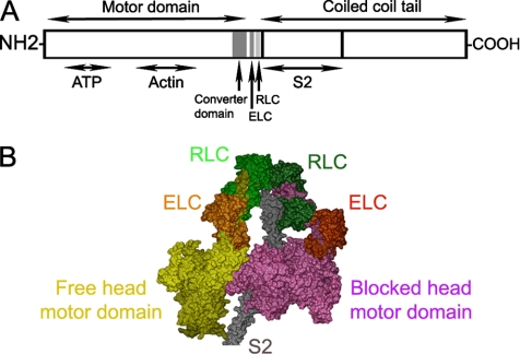
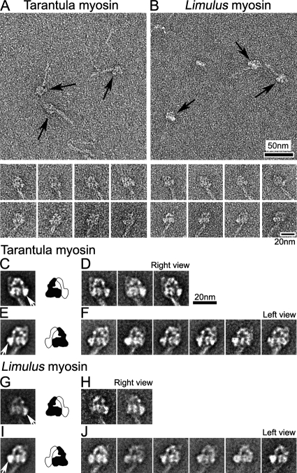
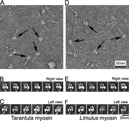
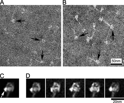
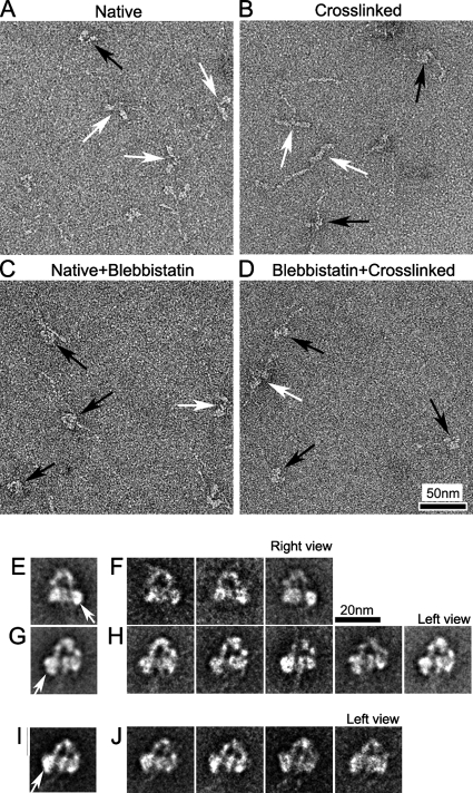
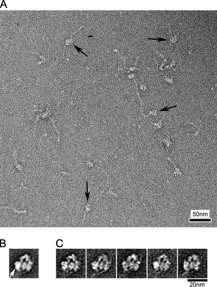
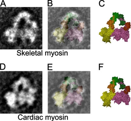
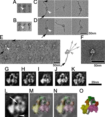
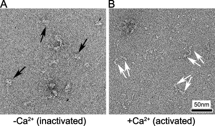
Intramolecular interaction between myosin heads, blocking key sites involved in actin-binding and ATPase activity, appears to be a critical mechanism for switching off vertebrate smooth-muscle myosin molecules, leading to relaxation. We have tested the hypothesis that this interaction is a general mechanism for switching off myosin II-based motile activity in both muscle and nonmuscle cells. Electron microscopic images of negatively stained myosin II molecules were analyzed by single particle image processing. Molecules from invertebrate striated muscles with phosphorylation-dependent regulation showed head-head interactions in the off-state similar to those in vertebrate smooth muscle. A similar structure was observed in nonmuscle myosin II (also phosphorylation-regulated). Surprisingly, myosins from vertebrate skeletal and cardiac muscle, which are not intrinsically regulated, undergo similar head-head interactions in relaxing conditions. In all of these myosins, we also observe conserved interactions between the 'blocked' myosin head and the myosin tail, which may contribute to the switched-off state. These results suggest that intramolecular head-head and head-tail interactions are a general mechanism both for inducing muscle relaxation and for switching off myosin II-based motile activity in nonmuscle cells. These interactions are broken when myosin is activated.


Figures

Figure 1.
Figure 1.
Structural organization of myosin II. (A) Schematic layout of myosin heavy chain, consisting of: motor domain (including converter domain and ATP- and actin-binding sites), light-chain domain (containing ELC and RLC binding sites), and the coiled-coil tail domain, including S2 at its N-terminal end. (B) Proposed model for head-S2 region of smooth-muscle myosin in the off-state (Blankenfeldt et al., 2006), built from atomic models of heads (Liu et al., 2003) and S2 (Blankenfeldt et al., 2006).
Figure 2.
Figure 2.
Compact head conformation of inactive (unphosphorylated) myosin molecules in relaxing conditions. (A and B) Fields (top panels) and galleries (bottom panels) of negatively stained tarantula and Limulus myosins. Arrows indicate head regions of compact molecules. (C–J) Global averages and selected class averages of tarantula and Limulus myosin images (alignment based on head features). (C and E) Global averages of tarantula myosin in right and left views, produced from 122 and 214 images, respectively. (G and I) Global averages of Limulus myosin in right and left views, from 52 and 267 images, respectively. (D, F, H, and J) Class averages, each based on 20–40 individual images in each orientation. Right and left views are defined according to position of the free head motor domain (white arrows in C, E, G, and I). Diagrams show interpretation of global averages in terms of blocked (black) and free (white) heads (see text). Scale bar, 20 nm in D applies to all average images.
Figure 3.
Figure 3.
Myosin conformation after glutaraldehyde cross-linking. (A and D) Fields of cross-linked tarantula and Limulus myosin molecules, respectively. Arrows indicate compact molecules. (B, C, E, and F) class averages of 20–40 images each of right and left views of tarantula and Limulus myosin. Total number of images used were 284 (B), 373 (C), 240 (E), and 303 (F).
Figure 4.
Figure 4.
Compact head conformation of unphosphorylated nonmuscle myosin IIA. (A and B) Fields of non-cross-linked and cross-linked nonmuscle myosin, respectively; arrows indicate compact heads. (C and D) Global and selected class averages (left view) from head-aligned images of cross-linked molecules (359 images total). Class averages contain 20–40 images each. White arrow indicates free head.
Figure 5.
Figure 5.
Structure of vertebrate skeletal myosin molecules after different chemical treatments under relaxing conditions. (A–D) Fields of (A) Native (no treatment), (B) Cross-linking (treated with 0.1% glutaraldehyde for 1 min), (C) Native+Blebbistatin (treated with 400 nM blebbistatin), and (D) Blebbistatin+Cross-linking (treated with 400 nM blebbistatin and then cross-linked). White and black arrows in fields indicate molecules with clearly separated or closely packed heads, respectively. (E and G) Global averages of blebbistatin-treated molecules. Right and left views from 65 and 125 images, respectively. (F and H) Selected class averages of blebbistatin-treated molecules, containing 20–40 images in each. (I and J) Global and selected class averages of 113 images of blebbistatin-treated and cross-linked molecules. Each class average contains 20–40 images. (E, G, and I) White arrows indicate free head motor domain. Scale bar, 20 nm in F applies to all average images.
Figure 6.
Figure 6.
Head–head interaction in vertebrate cardiac muscle myosin. (A) Field of blebbistatin-treated, cross-linked molecules; arrows indicate molecules with compact head appearance. (B and C) Global (B) and class averages (C) from 144 left view images. Each class average contains 20–40 images. White arrow indicates free head motor domain.
Figure 7.
Figure 7.
Fitting of interacting-head atomic model (PDB: 1i84) to vertebrate skeletal and cardiac myosin average images. (A) Selected average of blebbistatin-treated skeletal myosin (left view, from Figure 5H). (B) Superposition of equivalent view of atomic model (C; color scheme as in Figure 1) on A. (D) Global average of blebbistatin-treated and cross-linked cardiac myosin (left view, from Figure 6B). (E) Superposition of equivalent view of atomic model (F) on D.
Figure 8.
Figure 8.
Interaction between S2 and the blocked head. (A–D) Metal shadowed images of tarantula (A), Limulus (B), skeletal (C), and cardiac (D) myosins after cross-linking. (E and F) Gallery of non-cross-linked skeletal myosin molecules (E), together with their average (F). The first tail segment points down in all images (A–F). White arrowheads indicate point of emergence of S2 from the blocked head, and black arrowheads the tip of the tail. Black arrows in C–E point to the first bend in the tail. Diagram in E shows pathway of short (head to bend position) and long segments of tail. Note that long segment binds to blocked head in a position distinct from S2 (Burgess et al., 2007); C-terminal of tail is flexible beyond this point (gray arrowhead), with three different directions shown. (G–K) Average images of cross-linked myosins produced from tarantula (G; 20 images), Limulus (H; 16 images), nonmuscle (I; 20 images), skeletal (J; 36 images), and cardiac (K; 32 images). (L) Average image in the head region, produced from averaging images shown in G–K. (M) Superposition of (L) on equivalent left view of atomic model (PDB: 1i84; color scheme as in Figure 1). (N) Superposition of (L) on the atomic model with S2 (gray coiled-coil) included (O; Blankenfeldt et al., 2006). White arrow and arrowhead in L show S2 passing from head–head junction to blocked head and then emerging from blocked head, respectively. This is also seen in all individual average images (G–K).
Figure 9.
Figure 9.
Organization of myosin heads on activation. (A and B) Fields of scallop striated muscle myosin molecules in low- (A) and high- (B) calcium states. In A, black arrows show compact heads, whereas in B, paired white arrows point to separated pairs of heads in different molecules.

References

    1. Alberts B., Johnson A., Lewis J., Raff M., Roberts K., Walter P. Molecular Biology of the Cell. New York: Garland Science; 2007.
    1. Ankrett R. J., Rowe A. J., Cross R. A., Kendrick-Jones J., Bagshaw C. R. A folded (10 S) conformer of myosin from a striated muscle and its implications for regulation of ATPase activity. J. Mol. Biol. 1991;217:323–335. - PubMed
    1. Blankenfeldt W., Thoma N. H., Wray J. S., Gautel M., Schlichting I. Crystal structures of human cardiac beta-myosin II S2-Delta provide insight into the functional role of the S2 subfragment. Proc. Natl. Acad. Sci. USA. 2006;103:17713–17717. - PMC - PubMed
    1. Burgess S. A., Walker M. L., Thirumurugan K., Trinick J., Knight P. J. Use of negative stain and single-particle image processing to explore dynamic properties of flexible macromolecules. J. Struct. Biol. 2004;147:247–258. - PubMed
    1. Burgess S. A., Yu S., Walker M. L., Hawkins R. J., Chalovich J. M., Knight P. J. Structures of smooth muscle myosin and heavy meromyosin in the folded, shutdown state. J. Mol. Biol. 2007;372:1165–1178. - PubMed

Publication types

LinkOut - more resources