. 2008 Aug;19(8):3203-11.
doi: 10.1091/mbc.e07-08-0765. Epub 2008 May 21.

Elucidation of a TRPC6-TRPC5 channel cascade that restricts endothelial cell movement

Affiliations

Elucidation of a TRPC6-TRPC5 channel cascade that restricts endothelial cell movement

Pinaki Chaudhuri et al. Mol Biol Cell. 2008 Aug.

Abstract

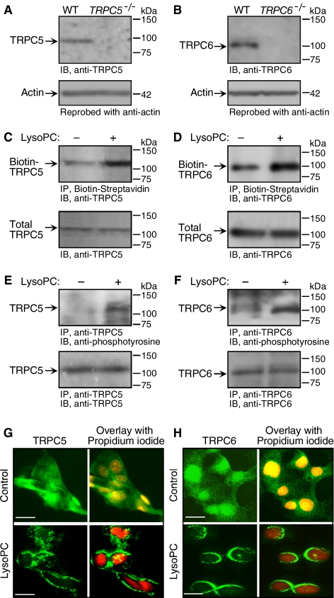
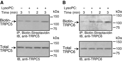
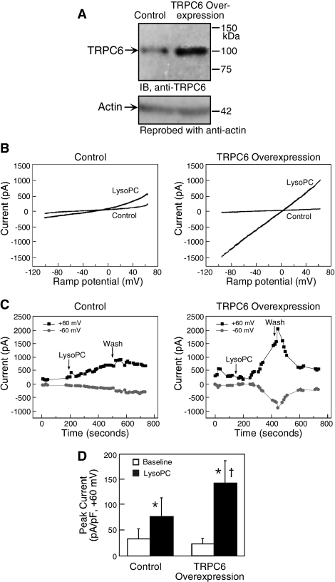
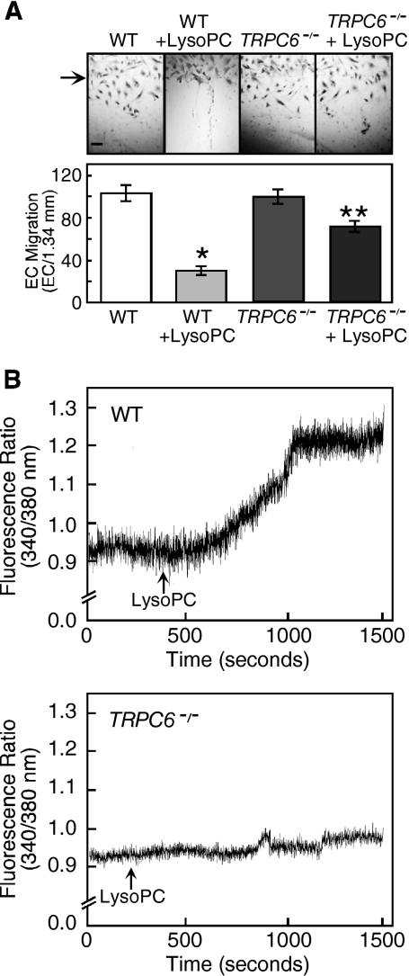
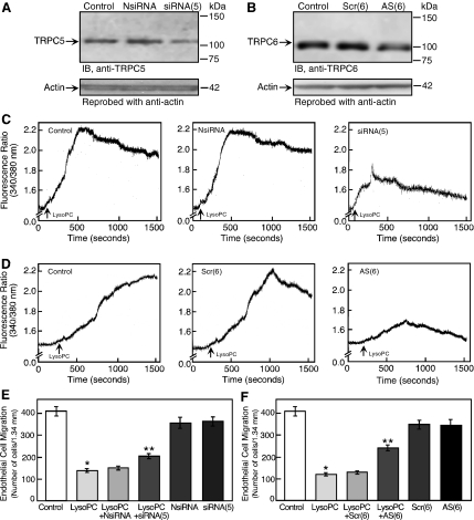
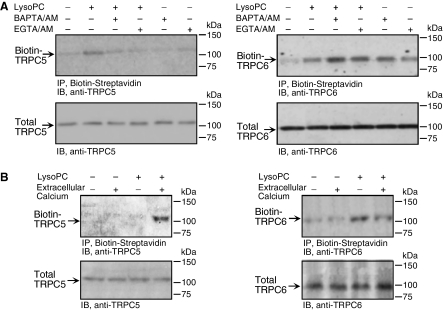
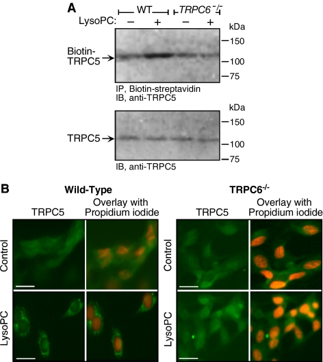
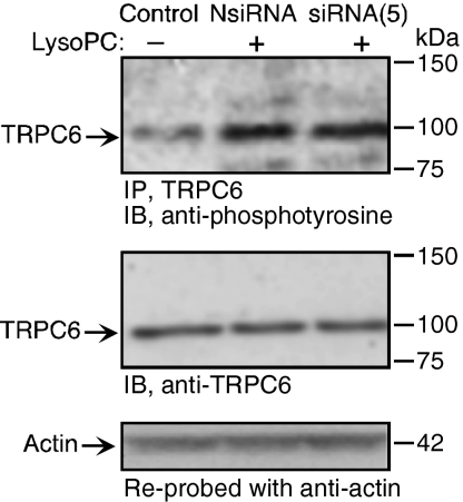
Canonical transient receptor potential (TRPC) channels are opened by classical signal transduction events initiated by receptor activation or depletion of intracellular calcium stores. Here, we report a novel mechanism for opening TRPC channels in which TRPC6 activation initiates a cascade resulting in TRPC5 translocation. When endothelial cells (ECs) are incubated in lysophosphatidylcholine (lysoPC), rapid translocation of TRPC6 initiates calcium influx that results in externalization of TRPC5. Activation of this TRPC6-5 cascade causes a prolonged increase in intracellular calcium concentration ([Ca(2+)](i)) that inhibits EC movement. When TRPC5 is down-regulated with siRNA, the lysoPC-induced rise in [Ca(2+)](i) is shortened and the inhibition of EC migration is lessened. When TRPC6 is down-regulated or EC from TRPC6(-/-) mice are studied, lysoPC has minimal effect on [Ca(2+)](i) and EC migration. In addition, TRPC5 is not externalized in response to lysoPC, supporting the dependence of TRPC5 translocation on the opening of TRPC6 channels. Activation of this novel TRPC channel cascade by lysoPC, resulting in the inhibition of EC migration, could adversely impact on EC healing in atherosclerotic arteries where lysoPC is abundant.


Figures

Figure 1.
Figure 1.
Translocation of TRPC5 and TRPC6 is induced by lysoPC. (A) Total protein was extracted from skeletal muscle tissues from wild-type (WT) and TRPC5−/− mice. Immunoblot analysis was performed to confirm the specificity of the anti-TRPC5 antibody. (B) Aortic ECs from wild-type and TRPC6−/− mice were lysed and immunoblot analysis was performed for total TRPC6 (n = 3) to confirm the specificity of the anti-TRPC6 antibody. (C) ECs were incubated with lysoPC for 1 h. Cell surface proteins were biotinylated, and immunoblot analysis performed for biotinylated TRPC5 (top panel). Before incubation with streptavidin-agarose beads, an aliquot of cell lysate was removed for immunoblot analysis to determine total TRPC5 (bottom panel). (D) Using similar methods, biotinylated TRPC6 (top panel) and total TRPC6 (bottom panel) were identified. (E) ECs were incubated with lysoPC for 1 h. TRPC5 was immunoprecipitated and immunoblot analysis was performed for phosphotyrosine (top panel) or total TRPC5 (bottom panel). (F) Using similar methods, tyrosine phosphorylated TRPC6 (top panel) and total TRPC6 (bottom panel) were identified. Representative blots of at least three separate experiments are shown in C–F. (G) At 60% confluence, ECs were made quiescent for 24 h, treated with or without lysoPC for 15 min, and then incubated with anti-TRPC5 antibody followed by Alexa 488–conjugated secondary antibody. TRPC5 location was assessed by fluorescence microscopy. Nuclei were detected by counterstaining with propidium iodide. (H) Using similar methods with anti-TRPC6 antibody, TRPC6 externalization was assessed by immunofluorescence microscopy. The images of TRPC5 (G) or TRPC6 (H) in ECs are the representative of four randomly chosen fields from each of three experiments. Original magnification, ×40. Bar, 40 μm.
Figure 2.
Figure 2.
TRPC6 is externalized more rapidly than TRPC5 after incubation with lysoPC. (A) ECs were grown to confluence in 10% FBS, washed, incubated with or without lysoPC for 1, 2, or 3 min. Cell surface proteins were biotinylated and immunoblot analysis performed for biotinylated TRPC5. Before incubation with streptavidin-agarose beads, an aliquot of cell lysate was removed for immunoblot analysis to determine total TRPC5. (B) Again using a biotinylation assay, the time course of TRPC6 externalization after addition of lysoPC was determined. Total TRPC6 was determined by immunoblot analysis. Representative blots of at least three separate experiments are shown in A and B.
Figure 3.
Figure 3.
Overexpression of TRPC6 amplifies the response of bovine aortic ECs to lysoPC. (A) ECs were transiently transfected with pcDNA3-TRPC6 for 24 h, made quiescent for an additional 24 h, and then lysed. Immunoblot analysis was performed for TRPC6 (top panel) and the blot was reprobed for actin to confirm equal loading (bottom panel). (B) For study of current–voltage relationships, ECs were transiently transfected with pcDNA3-TRPC6-eYFP or pcDNA-TRPC6 for 24 h and then made quiescent for 24 h. Ramp current traces (+60 to −100 mV over 125 ms) were obtained in whole cell patch-clamp studies using control ECs with empty expression vector pcDNA3 (left panel) and ECs overexpressing TRPC6 (right panel). Control trace represents the last trace before application of lysoPC. The bath solution was changed to one containing lysoPC (10 μM), and data collection was continued. (C) The time plots of current traces from the ECs shown in B are depicted for control ECs (left panel) and ECs overexpressing TRPC6 (right panel). After obtaining a stable baseline, the bath solution was changed to one containing 10 μM lysoPC (arrow). After ∼4 min, the bath solution was again changed to buffer without lysoPC (Wash), whereas data collection continued. (D) The peak current density (expressed as pico-amperes/pico-Faraday) was determined at baseline and after addition of lysoPC in five control ECs and seven ECs overexpressing TRPC6. The graph represents the mean ± SEM. * p < 0.05 compared with baseline and p = 0.07 compared with lysoPC-treated control ECs. (B and C) Representative of results obtained from five control ECs and seven ECs overexpressing TRPC6.
Figure 4.
Figure 4.
LysoPC has little effect on [Ca2+]i or migration of TRPC6−/− MAECs. (A) ECs were grown to confluence and made quiescent for 12 h, and then the migration assay was initiated in the presence or absence of lysoPC (10 μM). The arrow indicates the starting line of migration. Original magnification, ×40. Bar, 100 μm. The bottom panel depicts migration results represented as mean ± SD (n = 3; * p < 0.001 compared with WT control and ** p < 0.001 compared with TRPC6−/− control). (B) MAECs were loaded with fura 2-AM. After adjusting the baseline, lysoPC (10 μM) was added to cells (arrow), and relative change of [Ca2+]i was monitored in WT (top panel) and TRPC6−/− MAECs (bottom panel). Each trace is representative of four separate experiments.
Figure 5.
Figure 5.
TRPC5 or TRPC6 down-regulation blunted the lysoPC-induced [Ca2+]i rise and inhibition of migration. (A) For 24 h, ECs were incubated with negative control siRNA [NsiRNA] or siRNA targeted against TRPC5 mRNA [siRNA(5)]. ECs were then made quiescent for 24 h, lysed, and immunoblot analysis performed for TRPC5. Blots were reprobed for actin to confirm equal loading. (B) For 24 h, ECs were incubated with scrambled [Scr(6)] or antisense [AS(6)] oligonucleotide targeted against TRPC6 mRNA. Oligonucleotides were removed, and ECs were made quiescent for 24 h and then lysed, and immunoblot analysis was performed for TRPC6 (top panel). Blots were reprobed for actin to confirm equal loading (bottom panel). (C) ECs were transiently transfected for 24 h with NsiRNA or siRNA(5). The siRNA was removed and ECs were made quiescent for 12–24 h. Then ECs were loaded with fura 2-AM. After adjusting the baseline, lysoPC (12.5 μM) was added to cells (arrow), and relative change of [Ca2+]i was monitored in control cells (left panel), NsiRNA-transfected ECs (central panel), or siRNA(5)-transfected ECs (right panel). (D) Similarly, ECs were transiently transfected with Scr(6) or AS(6) oligonucleotides for 24 h, made quiescent, and then loaded with fura 2-AM. LysoPC (12.5 μM) was added (arrow), and relative change of [Ca2+]i was monitored in control cells (left panel), Scr(6)-transfected ECs (central panel), or AS(6)-transfected ECs (right panel). (E) ECs were transiently transfected for 24 h with NsiRNA or siRNA(5) and then made quiescent for 12 h. EC migration assay was initiated in the presence or absence of lysoPC (12.5 μM; n = 3). (F) ECs were transiently transfected with Scr(6) or AS(6) oligonucleotides for 24 h and then made quiescent for 12 h. EC migration assay was initiated in the presence or absence of lysoPC (12.5 μM; n = 4). In A–D, representative blots or traces of at least three separate experiments are shown. In E and F, results are represented as mean ± SD; *p < 0.001 compared with control and **p < 0.001 compared with lysoPC.
Figure 6.
Figure 6.
LysoPC-induced TRPC5 translocation is calcium-dependent, but TRPC6 translocation is calcium-independent. (A) ECs were preincubated with BAPTA/AM (25 μM) or EGTA/AM (30 μM) for 30 min before and during incubation with lysoPC (12.5 μM) for 3 min. Cell surface proteins were biotinylated, and immunoblot analysis was performed for biotinylated TRPC5 (left top panel) or TRPC6 (right top panel). Before incubation with streptavidin-agarose beads, an aliquot of cell lysate was removed for immunoblot analysis to determine total TRPC5 (left bottom panel) or TRPC6 (right bottom panel). (B) ECs were incubated with lysoPC (12.5 μM) for 30 min in the absence or presence of Ca2+ in KR buffer. Cell surface proteins were biotinylated, and immunoblot analysis was performed for biotinylated TRPC5 (left panel) or TRPC6 (right panel). Representative blots of five and three separate experiments are shown in A and B, respectively.
Figure 7.
Figure 7.
LysoPC-induced translocation of TRPC5 is dependent on TRPC6. (A) MAECs from wild-type (WT) and TRPC6−/− mice were incubated with lysoPC for 1 h. Cell surface proteins were biotinylated, and immunoblot analysis was performed for biotinylated TRPC5 (top panel). Before incubation with streptavidin-agarose beads, an aliquot of cell lysate was removed for analysis of total TRPC5 by immunoblot analysis (lower panel; n = 4). (B) WT and TRPC6−/− MAECs were grown to 60% confluency, washed, treated with or without lysoPC for 15 min, and then incubated with anti-TRPC5 antibody followed by Alexa 488–conjugated secondary antibody. TRPC5 location was assessed by immunofluorescence microscopy. Nuclei were detected by counterstaining with propidium iodide. The images of TRPC5 in ECs are the representative of four randomly chosen fields from each of three experiments. Original magnification, 40×. Bar, 40 μm.
Figure 8.
Figure 8.
LysoPC-induced phosphorylation of TRPC6 is not dependent on TRPC5. ECs were incubated with siRNA targeted against TRPC5 mRNA [siRNA(5)] or negative control siRNA [NsiRNA] for 24 h. siRNA was removed and after an additional 24 h, lysoPC (12.5 μM) was added for 1 h. Then TRPC6 was immunoprecipitated, and immunoblot analysis for phosphotyrosine was performed (top panel). Similarly treated or control ECs were lysed, and immunoblot analysis for TRPC6 was performed (middle panel). Blots were reprobed for actin to confirm equal loading (bottom panel). Representative blots of three separate experiments are shown.

Similar articles

Cited by

References

    1. Bezzerides V. J., Ramsey I. S., Kotecha S., Greka A., Clapham D. E. Rapid vesicular translocation and insertion of TRP channels. Nat. Cell Biol. 2004;6:709–720. - PubMed
    1. Boulay G., Zhu X., Peyton M., Jiang M., Hurst R., Stefani E., Birnbaumer L. Cloning and expression of a novel mammalian homolog of Drosophila Transient Receptor Potential (Trp) involved in calcium entry secondary to activation of receptors coupled by the Gq class of G protein. J. Biol. Chem. 1997;272:29672–29680. - PubMed
    1. Cayouette S., Lussier M. P., Mathieu E. -L., Bousquet S. M., Boulay G. Exocytotic insertion of TRPC6 channel into the plasma membrane upon Gq protein-coupled receptor activation. J. Biol. Chem. 2004;279:7241–7246. - PubMed
    1. Chaudhuri P., Colles S. M., Damron D. S., Graham L. M. Lysophosphatidylcholine inhibits endothelial cell migration by increasing intracellular calcium and activating calpain. Arterioscler. Thromb. Vasc. Biol. 2003;23:218–223. - PubMed
    1. Chaudhuri P., Colles S. M., Fox P. L., Graham L. M. Protein kinase Cδ-dependent phosphorylation of syndecan-4 regulates cell migration. Circ. Res. 2005;97:674–681. - PubMed

Publication types

LinkOut - more resources