

Optical coherence tomography of the cochlea in the porcine model
- PMID: 18496151
- PMCID: PMC2860954
- DOI: 10.1097/MLG.0b013e318173dd6c
Optical coherence tomography of the cochlea in the porcine model
Abstract
Objectives/hypothesis: To demonstrate the feasibility of optical coherence tomography in microstructural imaging of the porcine cochlea.
Study design: Ex vivo, porcine model.
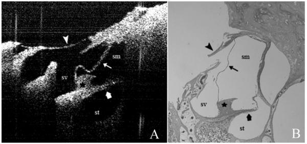
Methods: Optical coherence tomographic images of the porcine cochlea were obtained by thinning the bone from the basal turn of the cochlea leaving the endosteum intact. The images were compared with the corresponding histological sections.
Results: In the areas of thinned bone, images were obtained of the stria vascularis, Reissner's membrane, basilar membrane, tectorial membrane, scala media, scala tympani, and scala vestibuli. The bone was too thick for adequate light penetration in the areas where it was not thinned. Good histological correlation was obtained.
Conclusions: Cochlear and vestibular microanatomic structures of the pig cochlea were clearly identified with histological confirmation, suggesting the potential application of this noninvasive imaging modality for in vivo imaging of the human cochlea.
Figures
References
-
- Izatt JA, Hee MR, Swanson EA, et al. Micrometer-scale resolution imaging of the anterior eye in vivo with optical coherence tomography. Arch Ophthalmol. 1994;112:1584–1589. - PubMed
-
- Welzel J. Optical coherence tomography in dermatology: a review. Skin Res Technol. 2001;7:1–9. - PubMed
-
- Jang IK, Bouma BE, Kang DH, et al. Visualization of coronary atherosclerotic plaques in patients using optical coherence tomography: comparison with intravascular ultrasound. J Am Coll Cardiol. 2002;39:604–609. - PubMed
-
- Shen B, Zuccaro G., Jr Optical coherence tomography in the gastrointestinal tract. Gastrointest Endosc Clin North Am. 2004;14:551–571. - PubMed
Publication types
MeSH terms
Grants and funding
LinkOut - more resources
Full Text Sources
