. 2007 Mar;26(2):105-122.
doi: 10.1080/07352680701252817.

Multiple functions of DNA polymerases

Affiliations

Multiple functions of DNA polymerases

Miguel Garcia-Diaz et al. CRC Crit Rev Plant Sci. 2007 Mar.

Abstract

The primary role of DNA polymerases is to accurately and efficiently replicate the genome in order to ensure the maintenance of the genetic information and its faithful transmission through generations. This is not a simple task considering the size of the genome and its constant exposure to endogenous and environmental DNA damaging agents. Thus, a number of DNA repair pathways operate in cells to protect the integrity of the genome. In addition to their role in replication, DNA polymerases play a central role in most of these pathways. Given the multitude and the complexity of DNA transactions that depend on DNA polymerase activity, it is not surprising that cells in all organisms contain multiple highly specialized DNA polymerases, the majority of which have only recently been discovered. Five DNA polymerases are now recognized in Escherichia coli, 8 in Saccharomyces cerevisiae, and at least 15 in humans. While polymerases in bacteria, yeast and mammalian cells have been extensively studied much less is known about their counterparts in plants. For example, the plant model organism Arabidopsis thaliana is thought to contain 12 DNA polymerases, whose functions are mostly unknown. Here we review the properties and functions of DNA polymerases focusing on yeast and mammalian cells but paying special attention to the plant enzymes and the special circumstances of replication and repair in plant cells.


Figures
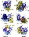
Figure 1
Figure 1. Structural similarity of DNA polymerases
Crystal structures of a representative member of each of the five DNA polymerase families (A, T. aquaticus Pol I; B, RB69 Pol; C, E. coli Pol III; X, Pol λ and Y, S. solfataricus Dpo4) and retrotranscriptases (RT). All DNA polymerases contain a general fold that can be likened to a right hand, with fingers, palm and thumb subdomains, colored in different shades of blue. Additional subdomains, colored yellow, are specific for each family or individual enzyme. Exo, 3′-5′ exonuclease domain; Nt, N-terminal domain; PHP, Polymerase and Histidinol Phosphatase domain; 8 kDa, 8 kDa domain; LF, Little Fingers domain; RH, RNAse H domain; CD, Connecting domain.
Figure 2
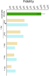
Figure 2. DNA synthesis fidelity
In vitro polymerization fidelity (defined as 1/error rate) of representative DNA polymerases. The fidelity for base substitutions (light brown bars) and insertion/deletion mutation (blue bars) is shown for of exo-deficient polymerases η (Matsuda, et al., 2001), κ (Ohashi, et al., 2000a) and λ (Bebenek, et al., 2003), for exo-proficient Pol ε and a Pol ε mutant deficient in proofreading (Shcherbakova, et al., 2003). An estimate of the error rate of the replication process in human cells (Drake, et al., 1998; Loeb, 2001) is shown in green. The error bar indicates the range of measured rates.
Figure 3
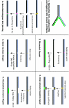
Figure 3. Different modes of synthesis during replication and repair
DNA polymerases carry out synthesis during many different processes. The requirements for the polymerase are very different in each pathway. Replication requires an extremely long, processive synthesis in the leading strand and shorter, interrupted patches of synthesis in the lagging strand. Different repair processes require patch synthesis anywhere from one to a few hundred nucleotides, and may also require synthesis on non-canonical substrates, such as DNA containing mismatches (shown in magenta) or different types of damage (yellow). The patch of synthesis performed by the polymerase is shown in green.

Similar articles

Cited by

References

    1. Abdulovic AL, Jinks-Robertson S. The in vivo characterization of translesion synthesis across UV-induced lesions in Saccharomyces cerevisiae: insights into Pol zeta- and Pol eta-dependent frameshift mutagenesis. Genetics. 2006;172:1487–1498. - PMC - PubMed
    1. Asturias FJ, Cheung IK, Sabouri N, Chilkova O, Wepplo D, Johansson E. Structure of Saccharomyces cerevisiae DNA polymerase epsilon by cryo-electron microscopy. Nat. Struct. Mol. Biol. 2006;13:35–43. - PubMed
    1. Autexier C, Lue NF. The Structure and Function of Telomerase Reverse Transcriptase. Annu. Rev. Biochem. 2006 - PubMed
    1. Bailey JC, 2nd, Heinhorst S, Cannon GC. Accuracy of Deoxynucleotide Incorporation by Soybean Chloroplast DNA Polymerases Is Independent of the Presence of a 3[prime] to 5[prime] Exonuclease. Plant Physiol. 1995;107:1277–1284. - PMC - PubMed
    1. Bailey S, Wing RA, Steitz TA. The structure of T. aquaticus DNA polymerase III is distinct from eukaryotic replicative DNA polymerases. Cell. 2006;126:893–904. - PubMed

LinkOut - more resources