Comparative Study
. 2008 Jul 20;509(3):239-58.
doi: 10.1002/cne.21690.

Developmental and comparative aspects of posterior medial thalamocortical innervation of the barrel cortex in mice and rats

Affiliations
Comparative Study

Developmental and comparative aspects of posterior medial thalamocortical innervation of the barrel cortex in mice and rats

Elizabeth A Kichula et al. J Comp Neurol. .

Abstract

The thalamocortical projection to the rodent barrel cortex consists of inputs from the ventral posterior medial (VPM) and posterior medial (POm) nuclei that terminate in largely nonoverlapping territories in and outside of layer IV. This projection in both rats and mice has been used extensively to study development and plasticity of highly organized synaptic circuits. Whereas the VPM pathway has been well characterized in both rats and mice, organization of the POm pathway has only been described in rats, and no studies have focused exclusively on the development of the POm projection. Here, using transport of Phaseolus vulgaris leucoagglutinin(PHA-L) or carbocyanine dyes, we characterize the POm thalamocortical innervation of adult mouse barrel cortex and describe its early postnatal development in both mice and rats. In adult mice, POm inputs form a dense plexus in layer Va that extends uniformly underneath layer IV barrels and septa. Innervation of layer IV is very sparse; a clear septal innervation pattern is evident only at the layer IV/Va border. This pattern differs subtly from that described previously in rats. Developmentally, in both species, POm axons are present in barrel cortex at birth. In mice, they occupy layer IV as it differentiates, whereas in rats, POm axons do not enter layer IV until 1-2 days after its emergence from the cortical plate. In both species, arbors undergo progressive and directed growth. However, no layer IV septal innervation pattern emerges until several days after the cytoarchitectonic appearance of barrels and well after the emergence of whisker-related clusters of VPM thalamocortical axons. The mature pattern resolves earlier in rats than in mice. Taken together, these data reveal anatomical differences between mice and rats in the development and organization of POm inputs to barrel cortex, with implications for species differences in the nature and plasticity of lemniscal and paralemniscal information processing.


Figures

Fig. 1
Fig. 1
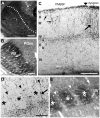
Laminar pattern of POm TCA innervation of adult mouse barrel cortex. A, B: Photomicrographs of adjacent coronal sections through somatosensory thalamus showing a representative injection of PHA-L (asterisk) in the POm nucleus (A). The image in (A) was taken in darkfield optics, so the injection site appears white against a dark background. The adjacent section (B) was processed histochemically for cytochrome oxidase activity to indicate nuclear borders. The dotted line in (A) corresponds to the border between VPM and POm nuclei shown in (B). C, D: Photomicrographs of coronal sections through adult mouse barrel cortex showing laminar innervation patterns of PHA-L-labeled POm TCAs and terminals (dark lines and stipple). C: Pia-to-white matter image through the border (vertical arrow) between the PMBSF and the dysgranular cortex (dysgran). In the PMBSF, labeled axons formed a dense terminal plexus in layer Va; innervation of layer IV was sparse. Some axons extended superficially, and branched obliquely to enter layer I (small arrows, left). Granular and supragranular layers of the dysgranular cortex were densely innervated (arrow, right). In this and subsequent figures, Roman numbers refer to cortical layers. D, E: higher power adjacent coronal sections through layer IV showing PHA-L-labeled axons and terminals (D) or cytochrome oxidase staining (E) to indicate position of intensely reactive barrels (asterisks) and more lightly stained, intervening septa (arrowheads). Septa are narrow in mice, and few axon terminals or fibers (arrow, D) were present in layer IV. Scale bars = 250 μm in A–C; 100 μm in D, E.
Fig. 2
Fig. 2
Septal-related patterning of POm TCAs in adult mouse PMBSF. Photomicrographs of adjacent sections cut in an oblique, tangential plane through middle layers of the PMBSF, showing PHA-L-labeled POm TCAs in darkfield optics (A), or cytochrome oxidase activity (B) which reveals borders between barrels (asterisks) and intervening septa. The plane of the oblique section extends from the upper half of layer IV (layer IV superficial) through the deeper half of layer IV (layer IV deep) into layer Va. PHA-L-labeled POm TCAs formed a dense plexus in layer Va that extended into the septa at the layer IV/Va border and into the deeper half of layer IV, but the density of fibers within the septa decreased significantly in passing into the superficial half of layer IV (arrows). Very few labeled axons were present within the barrels. The circles in the two images show matched positions of the same blood vessels. Scale bars = 100 μm in A, B.
Fig. 3
Fig. 3
Developmental progression of laminar innervation by POm TCAs in mouse barrel cortex. Pairs of confocal microscope images of coronal sections through developing mouse PMBSF at various postnatal ages from birth (P0) through P8 showing POm TCAs labeled by DiI (A,E,G,I,K) or DiD (C), and laminar cytoarchitecture revealed by Nissl counterstaining (B,D,F,H,J,L) of the corresponding dye-labeled sections. Dotted lines in the DiI/D images indicate borders between layers. A, B: At P0, many POm TCAs turned into barrel cortex from a subjacent stratum of labeled axons in the white matter (asterisk). Many axons penetrated the dense cortical plate (CP), and some were tipped by growth cones (inset, arrow; DiI-labeled axons are shown in white against the Nissl counterstaining). MZ, marginal zone. C, D: By P2, many radially oriented POm TCAs in layer V sent an oblique, horizontal branch into layer IV (arrows, C), which has differentiated from the CP by this age (D). E, F: By P4, overall density of labeled POm TCAs had increased in deep layers; long horizontal branches were evident in layer V (arrows, E). G, H: By P5, regularly-spaced, radially oriented branches spanned layer IV and extended into superficial layers, some of which (arrowheads, G) could be traced from long horizontal or oblique branches in layer V (arrows, G). I, J: By P6, the regularly spaced, radial branches that spanned layer IV (arrowheads, I) were evident; fewer labeled axons were found superficially in comparison with younger stages. Some oblique branches arose from layer V and traversed layer IV horizontally (arrows, I). K, L: By P8, features of an adult-like pattern emerged. Brackets (K) indicate position of image through layers IV/Va shown at higher power in inset. Regularly spaced, single fibers penetrated radially through layer IV in the PMBSF (arrowheads, inset). The layer Va plexus appeared scalloped at the border with layer IV. Adjacent to the PMBSF, labeled POm TCAs innervated densely all layers of dysgranular cortex (arrow, K). The cytoarchitectural border between the PMBSF and dysgranular cortex is shown by arrow in (L). Scale bars = 200 μm in B–L.
Fig. 3
Fig. 3
Developmental progression of laminar innervation by POm TCAs in mouse barrel cortex. Pairs of confocal microscope images of coronal sections through developing mouse PMBSF at various postnatal ages from birth (P0) through P8 showing POm TCAs labeled by DiI (A,E,G,I,K) or DiD (C), and laminar cytoarchitecture revealed by Nissl counterstaining (B,D,F,H,J,L) of the corresponding dye-labeled sections. Dotted lines in the DiI/D images indicate borders between layers. A, B: At P0, many POm TCAs turned into barrel cortex from a subjacent stratum of labeled axons in the white matter (asterisk). Many axons penetrated the dense cortical plate (CP), and some were tipped by growth cones (inset, arrow; DiI-labeled axons are shown in white against the Nissl counterstaining). MZ, marginal zone. C, D: By P2, many radially oriented POm TCAs in layer V sent an oblique, horizontal branch into layer IV (arrows, C), which has differentiated from the CP by this age (D). E, F: By P4, overall density of labeled POm TCAs had increased in deep layers; long horizontal branches were evident in layer V (arrows, E). G, H: By P5, regularly-spaced, radially oriented branches spanned layer IV and extended into superficial layers, some of which (arrowheads, G) could be traced from long horizontal or oblique branches in layer V (arrows, G). I, J: By P6, the regularly spaced, radial branches that spanned layer IV (arrowheads, I) were evident; fewer labeled axons were found superficially in comparison with younger stages. Some oblique branches arose from layer V and traversed layer IV horizontally (arrows, I). K, L: By P8, features of an adult-like pattern emerged. Brackets (K) indicate position of image through layers IV/Va shown at higher power in inset. Regularly spaced, single fibers penetrated radially through layer IV in the PMBSF (arrowheads, inset). The layer Va plexus appeared scalloped at the border with layer IV. Adjacent to the PMBSF, labeled POm TCAs innervated densely all layers of dysgranular cortex (arrow, K). The cytoarchitectural border between the PMBSF and dysgranular cortex is shown by arrow in (L). Scale bars = 200 μm in B–L.
Fig. 4
Fig. 4
Morphology of single POm TCA arbors in developing mouse barrel cortex. Representative examples of single, DiI-labeled POm TCAs in the PMBSF at P0 (A), P4 (B) and P8 (C). Individual arbors were selected from a total of 35 axons at P0 (n = 6 mice); from a total of 51 axons at P4 (n = 8 mice); and from a total of 43 axons at P8 (n = 9 mice). Scale bar = 100 μm in C (applies to A–C).
Fig. 5
Fig. 5
Features of the adult-like pattern of laminar innervation by POm TCAs emerge at the beginning of the second postnatal week in mice. Confocal images of carbocyanine dye-labeled POm TCAs in middle layers of PMBSF (A, B) or in dysgranular cortex (C). A: High-power image of a section through layers IV/Va taken from a P9 mouse. Dotted lines demarcate outlines of individual barrels; the barrels are visible by refringence. Septa are indicated by arrows. A few, radially oriented DiD-labeled axons entered or traversed layer IV through the septa (arrowheads); the origins of many could be identified as extensions from oblique or horizontal branches in the layer Va plexus. In this animal, the injection was small and very few axons were labeled, affording great clarity in the pattern. B: Image taken from a P10 mouse in which the DiI injection site was larger, resulting in a greater density of DiI-labeled POm TCAs. At this stage, the dense layer Va plexus was adult-like, as was the sparse innervation of layer IV. C: Image taken from same animal as that shown in (B); the POm TCA innervation of dysgranular cortex was dense and adult-like. Scale bars = 200 μm in A–C.
Fig. 6
Fig. 6
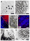
Emergence of a septal-related pattern of POm TCA innervation in developing mouse barrel cortex. A, B: Confocal microscope images of a tangential section through layer IV taken from a P5 mouse showing DiI-labeled POm TCAs (A) and barrel/septal cytoarchitecture revealed by Nissl-counterstaining (B). Asterisks show the same barrels in the two images; barrels are also visible in (A) by refringence. Short axon-fragments were found in both barrels (arrowhead) and septa; occasional longer, horizontally oriented axons were also seen in the septa (arrow). No overt septal pattern was yet evident at this age. C, D: A representative pair of adjacent, confocal images of coronal sections through the somatosensory thalamus of a P6 mouse in which two different carbocyanine dyes (C) were used simultaneously to label VPM (DiD, blue) and POm (DiI, red) TCAs. Nuclear borders were revealed cytoarchitecturally by Nissl staining (D); dotted lines delineate the extent of the VPM nucleus. E: Confocal image of a tangential section through layer IV taken from a mouse in which VPM and POm nuclei were both labeled as shown in (C). At P6, whisker-related clusters of VPM TCAs (blue) were well-formed as expected. Sparsely-distributed POm TCAs were found in both barrels and septa. F, G: Confocal images of an oblique, tangential section spanning the upper half of layer IV (upper right in the image) through layer Va (lower left in the image) taken from a P9 mouse showing DiI-labeled POm TCAs (F) and layer IV barrel/septal cytoarchitecture revealed by Nissl counterstaining (G). By this age, the dense layer Va plexus of POm TCAs was evident, as was a mature-like septal-pattern of innervation in the lower half of layer IV, particularly along barrel rows (arrowheads). The asterisks denote the position of the same, representative barrel in the two images. Scale bars = 200 μm in A, B, D–G (that in D also applies to C).
Fig. 7
Fig. 7
A mature-like septal-innervation pattern by POm TCAs is evident in mice by P10. Confocal images of an adjacent series of tangential sections spanning the full depth of layer IV (A–D) through the layer IV/Va border (E,F) taken from a P10 mouse showing DiD-labeled POm TCAs (A, C, E) and barrel/septal cytoarchitecture revealed by Nissl counterstaining (B, D, F). A mature-like pattern was fully resolved by this age. Labeled POm TCAs were very sparse in the upper half of layer IV (A, B), but in passing progressively deeper, they became increasingly concentrated in the septa, particularly along barrel rows (C,D). At the layer IV/Va border, the septal-innervation pattern was prominent, with labeled POm TCAs also appearing in the position of the barrels as the plane of the section dips into the layer Va plexus (E, F). Scale bars = 200 μm in A–F.
Fig. 8
Fig. 8
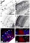
Developmental progression of laminar innervation by POm TCAs in rat barrel cortex. Confocal microscope images of coronal sections through developing rat PMBSF at various postnatal ages showing DiD-labeled POm TCAs (A,C) and corresponding laminar cytoarchitecture revealed by Nissl counterstaining (B,D), or showing dual labeling of VPM and POm TCAs (E–G). Dashed lines in the DiD images indicate borders between layers. A, B: At P0, POm TCAs entered barrel cortex from a white matter stratum of labeled axons (asterisk). Many axons were tipped by growth cones (arrowheads), but unlike mice, few penetrated the dense cortical plate (CP). Most axons were radially oriented; some however took long, horizontal or oblique trajectories into layer V (arrows). Small double arrows indicate a retrogradely-labeled corticothalamic cell. MZ, marginal zone. C, D: By P2, labeled axons remained mostly restricted to infragranular layers and avoided layer IV, which has emerged from the CP by this age. E: By P4, whisker-related clusters of VPM TCAs (DiD, blue) within layer IV barrels (asterisks) were well formed as expected. Branches of DiI-labeled POm TCAs (red) had by this age entered and extended through layer IV, although they occupied both septa and barrels (arrowheads). Other labeled POm TCAs took long, oblique trajectories through infragranular layers, spanning several barrel-widths (large arrows). Some radial axons extended into superficial layers (small double arrows), reaching layer I. F, G: Confocal images through middle layers taken from a P6 rat in which the VPM and POm TCA projections were labeled simultaneously. In (F), the channel showing DiD-labeled POm axons (red) is merged with that showing DiI-labeled VPM TCAs (blue), while in (G), the channel showing POm TCAs is displayed separately. Labeled POm TCAs remained in both septa (arrowhead) and barrels (arrows), but they appeared to be receding somewhat from barrels in comparison with younger ages. The dysgranular cortex was densely innervated by this age. Scale bars = 200 μm (A–G).
Fig. 9
Fig. 9
Morphology of single POm TCA arbors in developing rat barrel cortex. Representative examples of single, DiI-labeled POm TCAs in the PMBSF at P0 (A), P4 (B) and P8 (C). Individual arbors were selected from a total of 48 axons at P0 (n = 8 rats); from a total of 58 axons at P4 (n = 9 rats); and from a total of 38 axons at P8 (n = 7 rats). Scale bar = 100 μm in C (also applies to A–C).
Fig. 10
Fig. 10
Comparison between rats and mice in the extent of advancement of POm TCAs into barrel cortex during development. Graphs showing laminar distributions of single-axon endpoints (the most superficial point of each axon examined) plotted as a percentage of the total number of axons analyzed at P0 (A) and P4 (B) for both mice (black bars) and rats (gray bars). At P0, a substantial proportion of POm TCAs in mice penetrated the CP while none of the axons in our sample from rats did. A Fisher exact probability test showed that radial advancement of POm TCAs into the cortex of P0 mice was significantly different in comparison with that in P0 rats (p < 0.001). By P4, a greater proportion of rat POm TCAs extended into superficial layers in comparison with mice, and by this age, there were no significant differences in radial advancement of POm TCAs between species (p > 0.1). The numbers of axons and animals used for each species and age in this analysis are given in the legends to Figures 4 and 9.
Fig. 11
Fig. 11
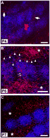
Emergence of septal-related pattern of POm TCA innervation in developing rat barrel cortex. Representative confocal microscope images of tangential sections through the middle of layer IV taken at three developmental ages from rats in which the VPM and POm TCA projections were labeled simultaneously by focal injection of DiD into the VPM nucleus (blue) and DiI into the POm nucleus (red). A: At P4, whisker-related clusters of VPM TCAs within barrels were well delineated as expected by this age. Labeled POm axons were sparse in layer IV, and were present in both barrels and septa (arrows). B: By P6, the density of POm TCA innervation in the septa had increased greatly (asterisks). Long, horizontally oriented POm TCAs were found along septa (arrowheads), or traversed barrels (small double arrows), spanning two rows. C: By P7, a mature-like septal-innervation pattern had resolved, in which the majority of labeled POm TCAs were restricted to the septa (asterisks). Scale bars = 200 μm in A–C.
Fig. 12
Fig. 12
Septa-related columns of POm TCA terminations spanning layers III through Va in rats. Photomicrographs of serially-ordered tangential sections spanning lower layer III (A, B), through layer IV (C, D) to layer Va (E, F) showing PHA-L-labeled POm TCAs (A, C, E) or cytochrome oxidase staining of the adjacent sections (B, D, F) to indicate the more intensely reactive barrels in layer IV (asterisks, D). PHA-L was injected into the POm nucleus on P6, the animal was killed on P8. Asterisks indicate the barrels in layer IV (C, D), or their corresponding positions in layer III (A, B) and layer Va (E, F). In layer IV, TCAs (arrows) are largely confined to septa. Labeled TCAs remain concentrated within septal-like columns in layer III and in layer Va, with fewer axons present above and below the position of the barrels (asterisks). The circle in the lower left corner of each image shows a blood vessel common to all sections. Scale bar = 200 μm in F (applies to A–F).
Fig. 13
Fig. 13
A schematic summary of the developmental sequence of POm TCA innervation of barrel cortex in mice (left column) and rats (right column). At P0, POm TCAs are present within the cortical layers in both species, but in mice, many more extend superficially, into the CP, than in rats, where they are mostly confined to deep layers. By P2, layer IV has emerged in both species; POm TCAs are present in layer IV in mice, but are mostly confined to infragranular layers in rats. By P4, barrel cytoarchitecture (dotted lines) and barrel-clusters of VPM TCAs (not shown) are both evident. In both species, POm axons penetrate layer IV and are present within barrels and septa; a greater proportion of POm TCAs in rats now extends superficially in comparison with mice. The major features of a mature-like pattern of POm TCA innervation emerge slightly earlier in rats (P5–8) in comparison with mice (P7–9). WM, white matter.

Similar articles

Cited by

References

    1. Abdel-Majid RM, Leong WL, Schalkwyk LC, Smallman DS, Wong ST, Storm DR, Fine A, Dobson MJ, Guernsey DL, Neumann PE. Loss of adenylyl cyclase I activity disrupts patterning of mouse somatosensory cortex. Nat Genet. 1998;19:289–291. - PubMed
    1. Agmon A, Yang LT, O’Dowd DK, Jones EG. Organized growth of thalamocortical axons from the deep tier of terminations into layer IV of developing mouse barrel cortex. J Neurosci. 1993;13:5365–5382. - PMC - PubMed
    1. Agmon A, Yang LT, Jones EG, O’Dowd DK. Topological precision in the thalamic projection to neonatal mouse barrel cortex. J Neurosci. 1995;15:549–561. - PMC - PubMed
    1. Ahissar E, Sosnik R, Haidarliu S. Transformation from temporal to rate coding in a somatosensory thalamocortical pathway. Nature. 2000;406:302–306. - PubMed
    1. Ahissar E, Sosnik R, Bagdasarian K, Haidarliu S. Temporal frequency of whisker movement. II. Laminar organization of cortical representations. J Neurophysiol. 2001;86:354–367. - PubMed

Publication types