. 2008 May 23;320(5879):1050-4.
doi: 10.1126/science.1158265.

Regulated protein denitrosylation by cytosolic and mitochondrial thioredoxins

Affiliations

Regulated protein denitrosylation by cytosolic and mitochondrial thioredoxins

Moran Benhar et al. Science. .

Abstract

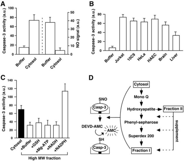
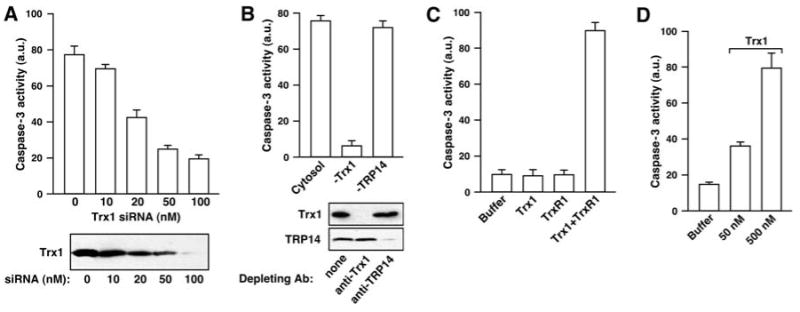
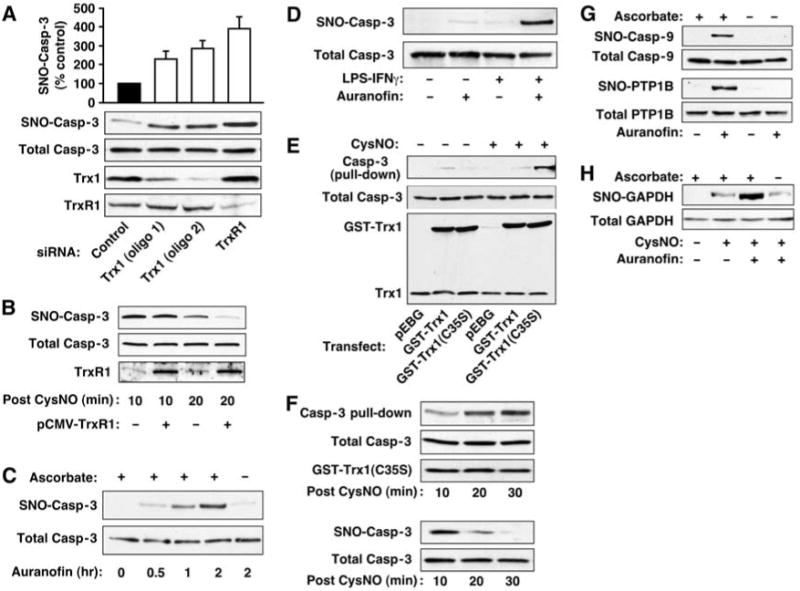
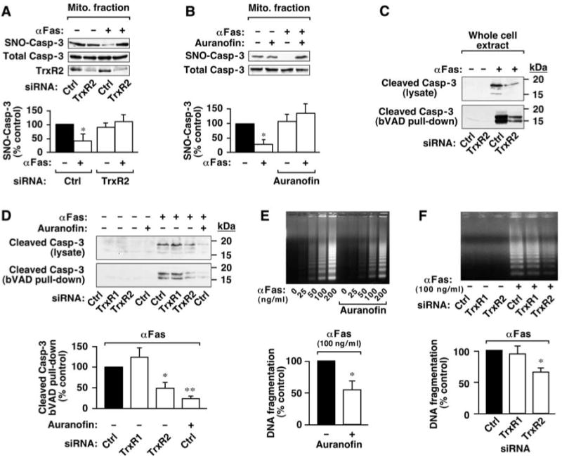
Nitric oxide acts substantially in cellular signal transduction through stimulus-coupled S-nitrosylation of cysteine residues. The mechanisms that might subserve protein denitrosylation in cellular signaling remain uncharacterized. Our search for denitrosylase activities focused on caspase-3, an exemplar of stimulus-dependent denitrosylation, and identified thioredoxin and thioredoxin reductase in a biochemical screen. In resting human lymphocytes, thioredoxin-1 actively denitrosylated cytosolic caspase-3 and thereby maintained a low steady-state amount of S-nitrosylation. Upon stimulation of Fas, thioredoxin-2 mediated denitrosylation of mitochondria-associated caspase-3, a process required for caspase-3 activation, and promoted apoptosis. Inhibition of thioredoxin-thioredoxin reductases enabled identification of additional substrates subject to endogenous S-nitrosylation. Thus, specific enzymatic mechanisms may regulate basal and stimulus-induced denitrosylation in mammalian cells.


Figures

Fig. 1
Fig. 1
Characterization of an enzymatic activity that denitrosylates SNO–caspase-3. Data in (A) to (C) are presented as mean ± SEM; n ≥ 3. (A) Reactivation of SNO–caspase-3 protease activity by cell cytosol. Immobilized SNO–caspase-3 (∼100 nM) was incubated for 30 min with a cytosolic extract prepared from Jurkat cells. (Left) Caspase activity was determined by using Z-DEVD-7-amino-4-methylcoumarin (DEVD-AMC). (Right) SNO content was assayed by using Hg-coupled photolysis-chemiluminescence. a.u., arbitrary unit. (B) Caspase activity after 30-min incubation of SNO–caspase-3 with cytosolic fractions (100 μg of protein) prepared from human cells or rat tissues. HAEC, primary human aortic endothelial cells. (C) SNO–caspase-3 was incubated with the cytosolic fraction from Jurkat cells or with cytosolic fractions after size-exclusion chromatography through Sephadex G-25 [high molecular weight (MW) fraction] and supplemented with GSH (0.5 mM), ATP (10 μM), NADH (10 μM), or NADPH (10 μM). (D) The procedure used to partially purify a SNO–caspase-3 denitrosylase (fig. S1, A and E, and table S1).
Fig. 2
Fig. 2
The Trx system is a major SNO–caspase-3 denitrosylating activity. Data are presented as mean ± SEM; n = 3. (A) Caspase-3 activity was determined (with Z-DEVD-AMC) after a 30-min incubation of SNO–caspase-3 (∼100 nM) with a cytosolic fraction prepared from untreated HeLa cells or from cells that were transfected for 3 days with siRNA for Trx1. (B) Caspase-3 activity was determined after a 30-min incubation of SNO–caspase-3 with HeLa cytosolic extract or cytosol that had been depleted of Trx1 or TRP14 by using specific antibodies against Trx1 or TRP14. (C) Caspase-3 activity was determined after a 30-min incubation of SNO–caspase-3 (∼100 nM) with NADPH (100 μM) and recombinant human Trx1 (10 nM) and/or recombinant rat TrxR1 (10 nM). (D) Caspase-3 activity after a 30-min incubation with recombinant Escherichia coli Trx1.
Fig. 3
Fig. 3
Trx1-TrxR1 mediates protein denitrosylation in vivo. Results are representative of three experiments. (A) 10C9 cells were transfected for 3 days with siRNA specific for Trx1 or TrxR1 or with control RNA. SNO–caspase-3 was assayed by biotin switch. The histogram summarizes results (mean ± SEM) of three experiments. (B) HEK cells were transfected for 24 hours with TrxR1 before treatment with CysNO (200 μM). Whole-cell extracts were prepared 10 or 20 min after CysNO exposure, and SNO–caspase-3 was assayed by biotin switch. (C) 10C9 cells were treated with auranofin (2 μM) for the times indicated, and SNO–caspase-3 was assayed by biotin switch. To verify biotin labeling specificity for SNO, the effect of omitting ascorbate in the assay is shown. (D) RAW264.7 cells were treated with auranofin (1 μM, 1 hour) or with LPS (1 μg/ml)–IFNγ (10 ng/ml) for 16 hours, or LPS-IFNγ plus auranofin (added for the last hour), and SNO–caspase-3 was assayed by biotin switch. (E) SNO-dependent interaction of Trx1 and caspase-3. HEK cells were co-transfected with caspase-3 and either GST-tagged wild-type Trx1, GST-tagged Trx1(C35S), or empty GST vector before treatment with CysNO (500 μM; 10 min). Affinity purification (pull-down) from lysates was with GSH-agarose. (F) HEK cells were treated as in (E) and lysed at different times after CysNO exposure. Pull-down of proteins was as in (E) (top). SNO–caspase-3 was assessed by biotin switch (bottom). (G and H) 10C9 cells were treated with auranofin (2 μM) or vehicle [dimethyl sulfoxide (DMSO)] for 2 hours, and S-nitrosylation of endogenous caspase-9, PTP1B, and GAPDH was assessed by biotin switch. For GAPDH (H), CysNO (500 μM; 20 min) was added after auranofin treatment.
Fig. 4
Fig. 4
The mitochondrial Trx system mediates Fas-induced denitrosylation of mitochondria-associated SNO–caspase-3 and promotes apoptotic signaling. (A) 10C9 cells were transfected for 3 days with siRNA for TrxR2 or with control RNA before exposure to CH11 monoclonal antibody against Fas (αFas; 50 ng/ml) for 2 hours. The amount of SNO–caspase-3 in a subcellular fraction enriched for mitochondria was evaluated by biotin switch. Results are mean ± SEM of four experiments. *P < 0.05 by analysis of variance (ANOVA). (B) 10C9 cells were left untreated or treated with auranofin for 1 hour, followed by Fas receptor stimulation and evaluation of mitochondrial SNO–caspase-3 as in (A). Results are the mean ± SEM of three experiments. *P < 0.05 by ANOVA. (C) 10C9 cells treated as in (A) were lysed in the presence of bVAD-FMK (10 μM), an affinity ligand for active caspase. Cleaved caspase-3 (<20 kD) was assessed by immunoblotting with antibodies against caspase-3 in lysates and after purification of active, bVAD-FMK–bound caspases with streptavidin-agarose (pull-down). Results are representative of three experiments. (D) 10C9 cells were transfected with siRNA specific for TrxR1 or TrxR2 or with control RNA. Cells were left untreated or treated with auranofin (1 μM; 1 hour) followed by treatment with anti-Fas (100 ng/ml; 2 hours). Caspase-3 cleavage and activation were assessed as in (C). Results (Fas-activated samples) are mean ± SEM of three experiments. *P < 0.05, **P < 0.01 by ANOVA. (E and F) 10C9 cells were treated with auranofin or transfected with siRNA specific for TrxR1 or TrxR2 as in (A) to (D). DNA fragmentation was assessed 6 hours after treatment with anti-Fas. Results (Fas-activated samples) are mean ± SEM of four experiments. *P < 0.05 by ANOVA.

Comment in

  • Biochemistry. SNO removal.
    Holmgren A. Holmgren A. Science. 2008 May 23;320(5879):1019-20. doi: 10.1126/science.1159246. Science. 2008. PMID: 18497281 No abstract available.

References

    1. Hess DT, Matsumoto A, Kim SO, Marshall HE, Stamler JS. Nat Rev Mol Cell Biol. 2005;6:150. - PubMed
    1. Mannick JB, et al. Science. 1999;284:651. - PubMed
    1. Erwin PA, Lin AJ, Golan DE, Michel T. J Biol Chem. 2005;280:19888. - PubMed
    1. Hoffmann J, Haendeler J, Zeiher AM, Dimmeler S. J Biol Chem. 2001;276:41383. - PubMed
    1. Kim JE, Tannenbaum SR. J Biol Chem. 2004;279:9758. - PubMed

Publication types

MeSH terms