. 2008 Jun 20;102(12):1512-9.
doi: 10.1161/CIRCRESAHA.108.172064. Epub 2008 May 22.

A specific CD36-dependent signaling pathway is required for platelet activation by oxidized low-density lipoprotein

Affiliations

A specific CD36-dependent signaling pathway is required for platelet activation by oxidized low-density lipoprotein

Kan Chen et al. Circ Res. .

Abstract

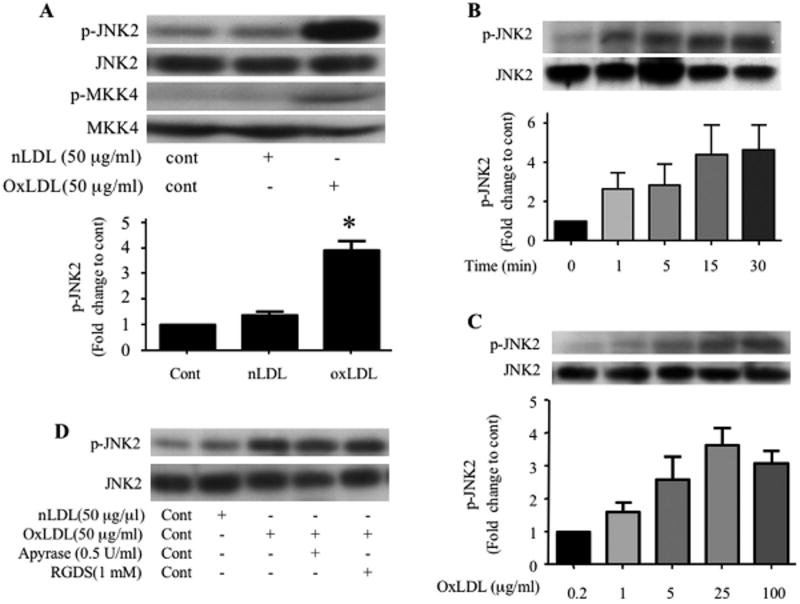
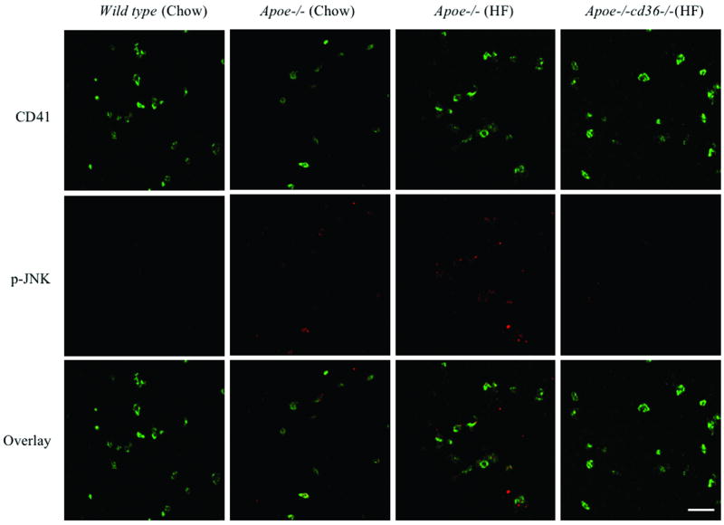
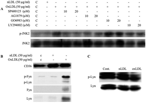
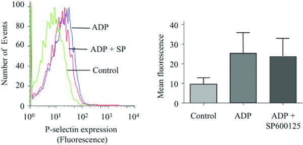
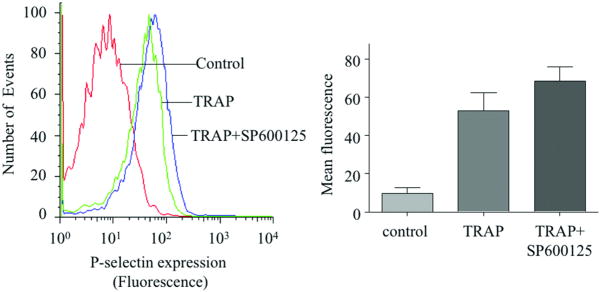
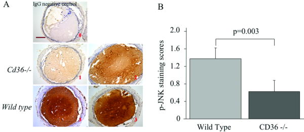
Platelet hyperactivity associated with hyperlipidemia may contribute to development of a prothrombotic state. We previously showed that oxidized low-density lipoprotein (oxLDL) formed in the setting of hyperlipidemia and atherosclerosis activated platelets in a CD36-dependent manner. We now show that mitogen-activated protein kinase c-Jun N-terminal kinase (JNK)2 and its upstream activator MKK4 were phosphorylated in platelets exposed to oxLDL. Using apoE(-/-) mice as a model of hyperlipidemia, we showed that JNK was constitutively phosphorylated in platelets in a CD36-dependent manner. Inhibition of src kinase activity reduced JNK phosphorylation by oxLDL. Immunoprecipitations revealed that active phosphorylated forms of src kinases Fyn and Lyn were recruited to CD36 in platelets exposed to oxLDL. Pharmacological inhibition of the mitogen-activated protein kinase JNK or src family kinases abolished platelet activation by oxLDL in vitro. Using a murine carotid artery thrombosis model we demonstrated CD36-dependent phosphorylation of platelet JNK within thrombi. Furthermore, pharmacological inhibition of JNK prolonged thrombosis times in wild-type but not cd36-null mice in vivo. These findings suggest that a specific CD36-dependent signaling pathway is required for platelet activation by oxLDL and may provide insights related to development of novel antiplatelet therapies more relevant to atherothrombosis than to normal hemostasis.


Conflict of interest statement

Disclosures: None.

Figures

Figure 1
Figure 1. OxLDL induces phosphorylation of JNK2 and MKK4 in platelets
Washed human platelets (2 × 10⁸/ml) containing 2 mM CaCl2 and 1 mM MgCl2 were incubated with native LDL or various concentrations of oxLDL over varying time points and then lysed. The lysates were analyzed by immunoblot with antibodies specific for phospho–JNK (p-JNK2, A, B, C), phospho-MKK4 (p-MKK4, A). The membranes were then stripped and re-probed with antibodies to the total relevant proteins to normalize the protein loaded. (D) Platelets were incubated with 50 μg/ml native LDL (Lane 2) or oxLDL with PBS (Lane 3), 0.5 U/ml apyrase (Lane 4) or 1 mM RGDS (Lane 5) for 5 minutes and then lysed. The lysates were analyzed by immunoblot as above for phospho-JNK (p-JNK) and total JNK. Results are representative of at least 3 independent experiments from different donors. The bar graph represents quantification of the phosphorylation of JNK2 (ratio of phosphorylated/total) expressed as relative values when compared with platelets without any treatment (A, B) or platelets treated with 0.2 μg/ml oxLDL (C). n=5 for A, n=3 for B and n=4 for C. c- control, nLDL- native LDL, oxLDL-oxidized LDL, * p < 0.05 when compared to control or nLDL treatment.
Figure 2
Figure 2. Basal phosphorylation of JNK is increased in resting platelets from hyperlipidemic mice in a CD36-dependent manner
(A) Platelets from WT and apoe-/- mice maintained on normal chow diet and apoe-/- mice and apoe-/-;cd36-/- mice maintained on high fat (HF) diet for 3 months at the age of 6 weeks were stained for phosphorylated JNK (Red), CD41 (Green) was stained simultaneously for platelet identification. Original magnification was 63×4 for all panels. Scale bar=20 μm. (B) Fluorescence intensity of 30 randomly selected platelets from 3 different mice (10 for each from 3 random fields) were determined with ImageJ software, and adjusted to the area of CD41 staining of the same platelet, used for p-JNK quantification. The bar graph represents Mean ± SE fluorescence values. (C) The lysates of platelets from apoe-/- mice maintained on normal chow diet and apoe-/- mice and apoe-/-;cd36-/- mice maintained on high fat (HF) diet for 3 months were analyzed by immunoblotting for the level of phospho-JNK as well as total JNK. The bar graph represents quantification of the phosphorylation of JNK (ratio of phosphorylated/total).
Figure 3
Figure 3. Recruitment of src family kinases to platelet CD36 is essential for oxLDL-mediated signaling
(A) Human platelets were incubated with the JNK inhibitor SP600125, src inhibitor AG1879, PKC inhibitor GO6983 or PI3K inhibitor LY294002 for 30 minutes prior to incubation with 50 μg/ml oxLDL. The platelet lysates were then analyzed by immunoblot as in figure 1 for JNK phosphorylation. (B) Platelets were incubated with oxLDL or native LDL and then lysed. CD36 was precipitated by FA6 anti-CD36 IgG. Precipitates were analyzed by immunoblot with antibodies to CD36, phospho-src (Y416), Fyn and Lyn. (C) Platelets were incubated with 50 μg/ml oxLDL or native LDL and then lysed. Lyn was immunoprecipitated from lysates and the precipitates were analyzed by immunoblot with a phosphotyrosine antibody (4G10) and Lyn antibody.
Figure 4
Figure 4. OxLDL-induced platelet activation of platelets is mediated by JNK and src kinases
(A-C) Human platelets were incubated with the specific JNK inhibitor SP600125 (final concentration of 20 uM) for 30 minutes prior to incubation with 50 μg/ml oxLDL (A) or 10 μM ADP (B) or 2 μM TRAP (SFLLRN) (C) and analyzed by flow cytometry with PE-labeled anti-P-selectin antibody. (D) Platelets were incubated with src family kinase inhibitor AG1879 (10 uM) for 30 minutes prior to incubation with 50 μg/ml oxLDL. Control platelets were treated with the vehicle, DMSO. Results are representative of at least 3 independent experiments from different donors. The bar graph represents mean fluorescence intensities measured with flow cytometry. The error bars are expressed as Mean ± SE. n=4 for A or n=3 for B, C, D.
Figure 5
Figure 5. JNK is phosphorylated during thrombus formation in a CD36-dependent manner
(A) Representative images of Immunohistochemical detection of phosphorylated JNK (p-JNK) in carotid thrombi from WT and cd36-/- mice. No staining with non-immune IgG control showed specificity. Brown indicated positive staining. Red font number indicates the score of the appropriate image. Scale bar=100 μm. (B) The bar graph represents Mean ± SE of staining score of 5 sections from 3 thrombi in each group.
Figure 6
Figure 6. JNK inhibition prolongs occlusion times in a CD36 dependent manner in vivo after carotid artery injury
Carotid thrombosis times were assessed by intravital video microscopy in irradiated thrombocytopenic WT and cd36-/- mice transfused with platelets from donor animals of identical genotype. Arteries were injured by topical application of FeCl3 (12.5%). Platelets were incubated with the JNK inhibitor SP600125 (400 nM) or vehicle control for 30 minutes prior to transfusion. Bar graph represents occlusion time. Data are expressed as Mean ± SE (n=5).

References

    1. Knowles DM, 2nd, Tolidjian B, Marboe C, D'Agati V, Grimes M, Chess L. Monoclonal anti-human monocyte antibodies OKM1 and OKM5 possess distinctive tissue distributions including differential reactivity with vascular endothelium. J Immunol. 1984;132:2170–2173. - PubMed
    1. Rhinehart-Jones T, Greenwalt DE. A detergent-sensitive 113-kDa conformer/complex of CD36 exists on the platelet surface. Archives of biochemistry and biophysics. 1996;326:115–118. - PubMed
    1. Abumrad NA, el-Maghrabi MR, Amri EZ, Lopez E, Grimaldi PA. Cloning of a rat adipocyte membrane protein implicated in binding or transport of long-chain fatty acids that is induced during preadipocyte differentiation. Homology with human CD36. The Journal of biological chemistry. 1993;268:17665–17668. - PubMed
    1. Van Nieuwenhoven FA, Verstijnen CP, Abumrad NA, Willemsen PH, Van Eys GJ, Van der Vusse GJ, Glatz JF. Putative membrane fatty acid translocase and cytoplasmic fatty acid-binding protein are co-expressed in rat heart and skeletal muscles. Biochemical and biophysical research communications. 1995;207:747–752. - PubMed
    1. Silverstein RL, Asch AS, Nachman RL. Glycoprotein IV mediates thrombospondin-dependent platelet-monocyte and platelet-U937 cell adhesion. The Journal of clinical investigation. 1989;84:546–552. - PMC - PubMed

Publication types

MeSH terms