. 2008 Jun 18;27(12):1736-46.
doi: 10.1038/emboj.2008.98. Epub 2008 May 22.

The iron-sulphur protein Ind1 is required for effective complex I assembly

Affiliations

The iron-sulphur protein Ind1 is required for effective complex I assembly

Katrine Bych et al. EMBO J. .

Abstract

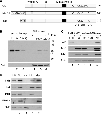
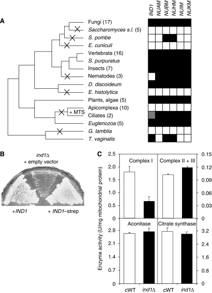
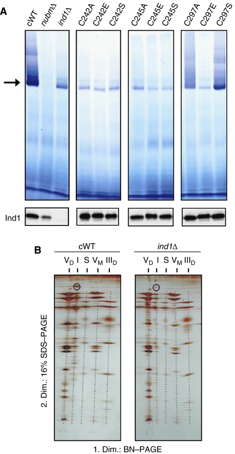
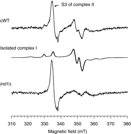
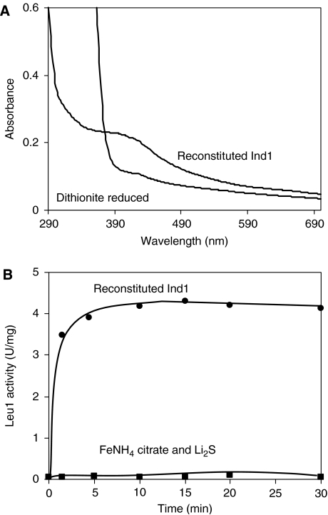
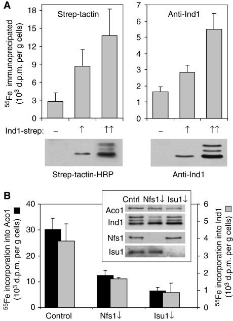
NADH:ubiquinone oxidoreductase (complex I) of the mitochondrial inner membrane is a multi-subunit protein complex containing eight iron-sulphur (Fe-S) clusters. Little is known about the assembly of complex I and its Fe-S clusters. Here, we report the identification of a mitochondrial protein with a nucleotide-binding domain, named Ind1, that is required specifically for the effective assembly of complex I. Deletion of the IND1 open reading frame in the yeast Yarrowia lipolytica carrying an internal alternative NADH dehydrogenase resulted in slower growth and strongly decreased complex I activity, whereas the activities of other mitochondrial Fe-S enzymes, including aconitase and succinate dehydrogenase, were not affected. Two-dimensional gel electrophoresis, in vitro activity tests and electron paramagnetic resonance signals of Fe-S clusters showed that only a minor fraction (approximately 20%) of complex I was assembled in the ind1 deletion mutant. Using in vivo and in vitro approaches, we found that Ind1 can bind a [4Fe-4S] cluster that was readily transferred to an acceptor Fe-S protein. Our data suggest that Ind1 facilitates the assembly of Fe-S cofactors and subunits of complex I.


Figures

Figure 1
Figure 1
Ind1 is targeted to mitochondria. (A) Cartoon of Mrp/NBP35-like proteins found in Yarrowia lipolytica. Cfd1, YALI0E19074g; Nbp35, YALI0E02354g; Ind1, YALI0B18590g. The proteins are 40% similar in amino-acid sequence but differ in their N termini. MTS, mitochondrial targeting sequence. Conserved cysteine motifs are indicated in black. Cys279 in Ind1 is drawn in grey, as it is not conserved in the mitochondrial group. (B) Polyclonal antibodies raised against recombinant Ind1–strep recognized a 30–31 kDa protein in cell extracts (20 μg protein per lane) of Y. lipolytica expressing full-length IND1 or IND1–strep (IND1s) under the control of its own promoter from plasmid pUB4. (C) Immunoblot showing the mitochondrial localization of Ind1. Y. lipolytica cells expressing IND1–strep were treated with zymolyase, broken in a Dounce homogenizer (Tot=total lysate) and fractionated in mitochondria (Mit) and post-mitochondrial supernatant (PMS). Protein (20 μg) was separated by SDS–PAGE, blotted and labelled with antibodies against Ind1, the mitochondrial proteins aconitase (Aco1) or cysteine desulphurase (Nfs1) or cytosolic actin. (D) Immunodetection of Ind1 in the mitochondrial matrix, associated with membranes. Mitochondria (50 μg protein) of Y. lipolytica expressing IND1–strep were incubated in hypotonic buffer to swell the organelles and break the outer membrane, followed by 10 min incubation and centrifugation in the presence of 150 mM KCl to separate soluble and membrane-bound proteins of the intermembrane space (Ims) from mitoplasts (Mp). The pellet (Mp) was resuspended in hypotonic buffer and subjected to three rounds of freeze–thawing, followed by centrifugation to separate soluble matrix proteins (Mtx) and membranes (Mem). The volume of each mitochondrial fraction was adjusted to 50 μl in 1 × gel loading buffer, and 20 μl of each fraction was analysed by SDS–PAGE and immunoblotting to visualize Ind1, Nfs1, the Fe–S scaffold protein Isu1 (soluble matrix protein), Rieske Fe–S protein (integral membrane protein of complex III) and cytochrome c (Cytc; protein of the intermembrane space). Note that the separation of cytochrome c from the mitoplasts was incomplete in this experiment. Similar results were obtained for cells expressing Ind1 without Strep tag (not shown).
Figure 2
Figure 2
IND1 is phylogenetically and functionally linked to complex I. (A) Coevolution of IND1 and genes for Fe–S protein subunits of complex I. A cross indicates the loss of IND1 in that lineage, and +MTS indicates the gain of a mitochondrial targeting signal in an IND1 paralogue. White and black squares represent the absence or presence, respectively, of the gene indicated above each column. Grey indicates the presence of an IND1 paralogue with a mitochondrial targeting signal. Abbreviated species names: Saccharomyces s.l. Saccharomyces sensu lato; S. (Schizosaccharomyces) pombe; E. (Encephalitozoon) cuniculi; S. (Strongylocentrotus) purpuratus; D. (Dictyostelium) discoideum; E. (Entamoeba) histolytica; G. (Giardia) lamblia; T. (Trichomonas) vaginalis. (B) Growth of Y. lipolytica strain ind1Δ in which the IND1 gene was deleted (centre, transformed with an empty plasmid) and the ind1Δ strain expressing the wild-type IND1 gene (left), or IND1 fused to the Strep tag coding sequence (right), expressed under the control of the IND1 promoter from the pUB4 plasmid. (C) Activities of Fe–S enzymes in purified mitochondria from ind1Δ cells (black bars) or the IND1-complemented wild type (cWT, white bars). Complementation with IND1 or IND1–strep gave similar results (not shown). Complex I was measured as NADH:HAR oxidoreductase activity in alamethicin-permeabilized mitochondria. Complex II plus III activity was measured following the electron transfer from succinate to cytochrome c in intact mitochondria. Aconitase activity was assayed following cis-aconitate consumption. Citrate synthase activity served as a non-Fe–S enzyme control. Error bars represent the standard deviation, n=3.
Figure 3
Figure 3
Deletion of IND1 leads to a major decrease in fully assembled complex I. (A) In-gel staining of complex I in mitochondrial membranes (upper panels) and immunostaining of Ind1 protein (lower panels). Mitochondrial membrane proteins were separated by BN–PAGE and incubated with NADH and NBT to visualize NADH dehydrogenase activity. Monomeric complex I (with a molecular mass of 947 kDa) is marked by an arrow. Activity-stained bands of higher molecular mass represent supercomplexes containing complex I. cWT (ind1Δ+pUB4-IND1); nubmΔ, a strain lacking the gene encoding the 51-kDa NUBM subunit of complex I; ind1Δ carrying the pUB4 plasmid; C242A–C279S, point mutations that exchange cysteines at positions 242, 245 or 279 in pUB4-IND1. (B) Two-dimensional (BN/SDS) PAGE analysis of mitochondrial membranes from complemented wild-type (cWT) and ind1Δ cells, stained with silver. The following multiprotein assemblies are indicated by dashed grey lines: VD and VM, dimeric and monomeric forms of complex V; I, complex I; S, an incompletely characterized supercomplex that contains complex III, IIID, dimeric form of complex III. The 75-kDa NUAM subunit of complex I is circled.
Figure 4
Figure 4
Deletion of IND1 results in significantly diminished EPR signals of complex I. EPR spectra of mitochondrial membranes from complemented wild type (cWT, top) and ind1Δ cells (bottom). A spectrum of isolated complex I is shown for comparison (middle). Samples were treated with 2 mM NADH, frozen and EPR spectra were recorded at 12 K. Instrument parameters: microwave frequency 9.47 GHz, modulation amplitude 0.64 mT, modulation frequency 100 kHz, microwave power 5 mW.
Figure 5
Figure 5
Ind1 binds a labile Fe–S cluster in vitro. (A) Binding of an [4Fe–4S] cluster to Ind1 in vitro as revealed by UV–Vis spectroscopy. Recombinant Ind1–strep (0.85 mg/ml, 26 μM) was reconstituted and desalted as described in Materials and methods. UV–Vis spectra were recorded in 100 mM Tris–HCl pH 9.0, 100 mM NaCl in the presence and absence of 2 mM sodium dithionite (as indicated). (B) Ind1 can efficiently transfer an Fe–S cluster to a model substrate, apo-isopropylmalate isomerase (Leu1). Reconstituted Ind1 (4 μM) was mixed with reduced apo-Leu1 (4 μM) under anaerobic conditions and Leu1 enzyme activity was measured at regular intervals (circles). As a control, activation of 4 μM apo-Leu1 with ferric ammonium citrate and Li2S at concentrations identical to those present in Ind1 was carried out (squares).
Figure 6
Figure 6
Ind1 binds an Fe–S cluster in vivo. (A) Wild-type S. cerevisiae cells carrying an empty plasmid (−) or plasmids for intermediate (↑) or high (↑↑) expression of Y. lipolytica IND1–strep were radiolabelled with 55Fe. Cells were harvested after 2 h and, following preparation of a cell extract, Ind1–strep was immunoprecipitated with either Strep-Tactin or Ind1 antibodies coupled to sepharose A. The 55Fe associated with the protein was quantified by scintillation counting. The expression levels of Ind1–strep were confirmed by immunoblotting (lower panels). (B) Iron-55 labelling and immunoprecipitation of Ind1 or endogenous aconitase (Aco1) upon depletion of the cysteine desulphurase Nfs1 (Nfs1↓) or the scaffold protein Isu1 (Isu1↓). Wild-type S. cerevisiae (control), Gal-NFS1 or Gal-ISU1/isu2Δ strains were transformed with a plasmid to overexpress Y. lipolytica IND1–strep, and grown on glucose to downregulate the expression of NFS1 or ISU1 as indicated. Expression of Aco1, Ind1, Nfs1 and Isu1 was visualized by immunoblotting (inset). The scintillation values were corrected for the average background signal obtained from experiments with cells containing an empty plasmid (for Ind1; 1.6 × 10³ d.p.m./g cells, see A), or with aco1Δ cells (for aconitase; 1.3 × 10³ d.p.m./g cells). Error bars represent the standard deviation, n=3–5.

References

    1. Abdrakhmanova A, Dobrynin K, Zwicker K, Kerscher S, Brandt U (2006) Functional sulfurtransferase is associated with mitochondrial complex I from Yarrowia lipolytica, but is not required for assembly of its iron–sulfur clusters. FEBS Lett 579: 6781–6785 - PubMed
    1. Balk J, Lobréaux S (2005) Biogenesis of iron–sulfur proteins in plants. Trends Plant Sci 10: 324–331 - PubMed
    1. Beinert H (2000) Iron–sulfur proteins: ancient structures, still full of surprises. J Biol Inorg Chem 5: 2–15 - PubMed
    1. Brandt U (2006) Energy converting NADH:quinone oxidoreductase (complex I). Annu Rev Biochem 75: 69–92 - PubMed
    1. Dunning CJR, McKenzie M, Sugiana C, Lazarou M, Silke J, Connelly A, Fletscher JM, Kirby DM, Thorburn DR, Ryan MT (2007) Human CIA30 is involved in the early assembly of mitochondrial complex I and mutations in its gene cause disease. EMBO J 26: 3227–3237 - PMC - PubMed

Publication types

MeSH terms

LinkOut - more resources