. 2008 Jun 18;27(12):1694-705.
doi: 10.1038/emboj.2008.94. Epub 2008 May 22.

Alu element-mediated gene silencing

Affiliations

Alu element-mediated gene silencing

Ling-Ling Chen et al. EMBO J. .

Abstract

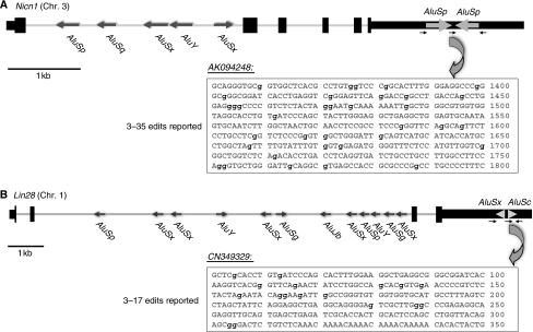
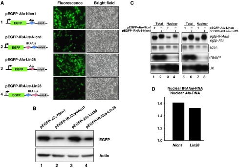
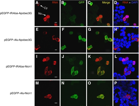
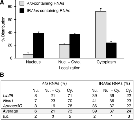
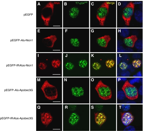
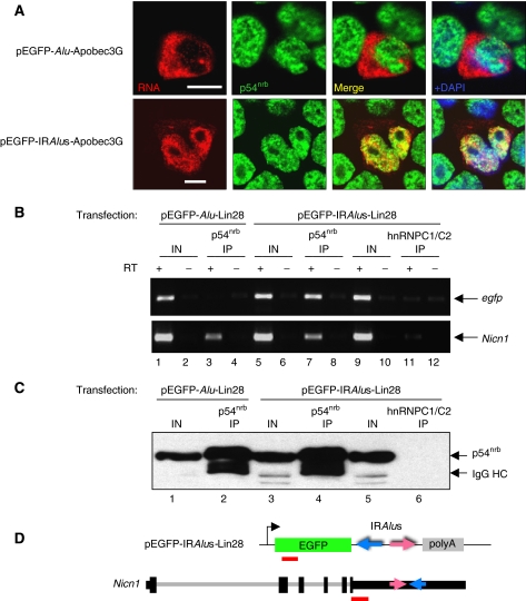
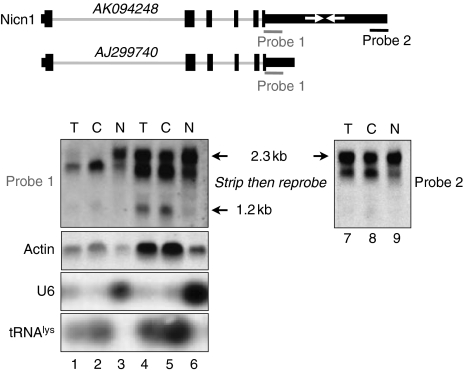
The Alu elements are conserved approximately 300-nucleotide-long repeat sequences that belong to the SINE family of retrotransposons found abundantly in primate genomes. Pairs of inverted Alu repeats in RNA can form duplex structures that lead to hyperediting by the ADAR enzymes, and at least 333 human genes contain such repeats in their 3'-UTRs. Here, we show that a pair of inverted Alus placed within the 3'-UTR of egfp reporter mRNA strongly represses EGFP expression, whereas a single Alu has little or no effect. Importantly, the observed silencing correlates with A-to-I RNA editing, nuclear retention of the mRNA and its association with the protein p54(nrb). Further, we show that inverted Alu elements can act in a similar fashion in their natural chromosomal context to silence the adjoining gene. For example, the Nicolin 1 gene expresses multiple mRNA isoforms differing in the 3'-UTR. One isoform that contains the inverted repeat is retained in the nucleus, whereas another lacking these sequences is exported to the cytoplasm. Taken together, these results support a novel role for Alu elements in human gene regulation.


Figures

Figure 1
Figure 1
Organization and Alu characterization of Nicn1 and Lin28. The genomic sequences of Nicn1 (A) and Lin28 (B) are drawn to scale. Exons and UTRs are shown as black bars, with coding regions being thicker. The Alu elements present in these two genes are shown as gray arrows with the indicated orientations. There is a single pair of IRAlus in each of the 3′-UTRs of Nicn1 and Lin28. The small black arrows indicate the PCR primers, their directions and their relative positions for the cloning sequences on the pEGFP-C1 vector. AK094248 shows one of the highly edited mRNA sequences of Nicn1 in the UCSC genome browser, whereas CN349329 shows one of the highly edited mRNA sequences of Lin28. The edited residues are denoted in lowercase in bold.
Figure 2
Figure 2
IRAlus in the 3′-UTR of egfp mRNA suppress EGFP expression at a post-transcriptional level. (A) IRAlus in the 3′-UTR of egfp mRNA suppress EGFP expression. IRAlus and Alu were PCR-amplified from the 3′-UTR of either Nicn1 or Lin28 and then inserted separately into the 3′-UTR of egfp mRNA. HEK293 cells were transfected with the indicated plasmids and fluorescence pictures were taken 44 h after transfection. (B) The expression of EGFP from the same batch of transfected HEK293 cells as described in panel A was investigated by western blotting, by probing with anti-GFP antibody. Actin was used as the loading control. (C) IRAlus RNAs are retained in the nucleus. Total and nuclear RNAs were isolated from the same batch of transfected HEK293 cells used in panels A and B and then resolved on a denaturing agarose gel. Transcripts of egfp-tagged RNAs were probed with a Dig-labelled egfp fragment. Actin RNA was used as the loading control; tRNAlys and U6 snRNA were used as markers for nuclear/cytoplasmic RNA isolation. (D) Preferential retention of IRAlus RNAs within the nucleus compared with single Alu peers. Total and nuclear IRAlus RNAs, as well as Alu-containing RNAs, were quantified from panel C and normalized to the relative amount of actin mRNAs. The ratio was obtained by comparison of the normalized value of the nuclear-retained IRAlus RNA to those of the nuclear-retained Alu RNA.
Figure 3
Figure 3
Nuclear retention of IRAlus-containing RNAs correlates with silencing of EGFP expression. RNA in situ hybridization (A, E, I, M) was performed with Dig-labelled antisense egfp probe (red) for each different transfection with plasmids encoding either IRAlus or Alu-RNA, and representative images are shown. No signals were detected with the Dig-labelled sense strand egfp fragment (data not shown). EGFP was visualized using anti-GFP antibodies (B, F, J, N). The white arrows in panel A identify cells in which mRNA is in either the cytoplasm or nucleus. Note that when RNA expression in the cytoplasm is higher, in these cells the expression of GFP is also higher. When the RNA is retained in the nucleus, GFP expression is reduced. Panels C, G, K and O merge the RNA and GFP signals, and panels D, H, L and P merge the RNA signal with nuclear DAPI staining. Scale bars 10 μm.
Figure 4
Figure 4
Subcellular distribution of IRAlus RNA. HEK293 cells were transfected with the plasmids described in Figures 2 and 3 and RNA in situ hybridization was performed with a Dig-labelled antisense egfp probe as in Figure 3. (A) A total of 200 transfected cells were recorded randomly by confocal microscopy following each different transfection, and the percentage of each distinct localization pattern of IRAlus-RNA or Alu-RNA was recorded. The average percentage of each pattern was calculated by the mean of IRAlus or Alu-RNA that comes from different genomic locus (Nicn1, Lin28 and Apobec3G). Signals that were exclusively nuclear are labelled ‘Nucleus'. Cells with RNA distributed both in the nucleus and cytoplasm are labelled ‘Nuc.+Cyto'. Cells with exclusively cytoplasmic signals are labelled ‘Cytoplasm'. The criteria used for assignment are illustrated in Supplementary Figure 3. The results are graphed as the sum of results from the various plasmids. (B) Overall results and statistical analysis of distribution patterns are tabulated. s.d., standard deviation.
Figure 5
Figure 5
IRAlus-RNA is retained in the nucleus and colocalizes with T7-p54nrb. RNA in situ hybridization was performed with Dig-labelled antisense egfp probe (red) for each different transfection with plasmids encoding either IRAlus or Alu-RNA, and representative images are shown (A, E, I, M, Q). No signals were detected with the Dig-labelled sense strand egfp fragment (data not shown). Co-transfected T7-p54nrb was visualized with anti-T7 antibody (green; B, F, J, N, R). Panels C, G, K, O and S merge the RNA and T7-p54nrb signals, and panels D, H, L, P and T merge the RNA and protein signals with nuclear DAPI staining. Scale bars, 10 μm.
Figure 6
Figure 6
IRAlus RNA and endogenous Nicn1 associate with p54nrb. (A) Colocalization of IRAlus RNA and endogenous p54nrb. HEK293 cells were transfected with plasmids encoding Alu RNA (upper panel) and IRAlus RNA (lower panel), and RNA in situ hybridization was carried out with Dig-labelled antisense egfp probe (red). Endogenous p54nrb was visualized with anti-p54nrb antibody (green). Nuclei were stained with DAPI. Scale bars, 10 μm. (B) IP from pEGFP-Alu-Lin28- or pEGFP-IRAlus-Lin28-transfected HEK293 cells using anti-p54nrb antibody or anti-hnRNPC1/C2 antibody (mock IP). RT–PCR of egfp from the IP using hexamer primers showed amplification only in the pEGFP-IRAlus-Lin28-transfected cells by anti-p54nrb IP, but not by IP with anti-hnRNPC1/C2 antibody or by IP in the pEGFP-Alu-Lin28-transfected cells by anti-p54nrb IP (upper panel). Endogenous Nicn1 RNA was associated with p54nrb in both transfections, but did not associate with hnRNP C1/C2 (lower panel). (C) Western blotting using anti-p54nrb antibody was used with extracts from the sample in panel B to confirm the specificity of IP. (D) Schematic representation of the fragments (red bars) in the pEGFP-C1 vector and the endogenous Nicn1 that were analysed by PCR amplification in panel B.
Figure 7
Figure 7
Characterization of Nicn1. Northern blot analysis of Nicn1 RNA in nuclear and cytoplasmic fractions from HEK293 cells revealed that a 1.2 kb band was enriched in the cytoplasmic fraction. Two probes from the 3′-UTR of Nicn1 were used for the northern blot. Probe 1 (red bar) is located upstream of the IRAlus whereas probe 2 (green bar) is located downstream of IRAlus. tRNAlys and U6 were used as makers for the nuclear and cytoplasmic RNA fractionation. In the bottom panel, total (T), cytoplasmic (C) and nuclear (N) RNAs were analysed by northern blotting. Lanes 1–3 are the same samples as lanes 4–6, except that a less amount of the samples was loaded onto the gel. Following hybridization with probe 1, the membrane representing lanes 4–6 was stripped and re-probed with probe 2 (lanes 7–9).

References

    1. Ambros V, Horvitz HR (1984) Heterochronic mutants of the nematode Caenorhabditis elegans. Science 226: 409–416 - PubMed
    1. Athanasiadis A, Rich A, Maas S (2004) Widespread A-to-I RNA editing of Alu-containing mRNAs in the human transcriptome. PLoS Biol 2: e391. - PMC - PubMed
    1. Backofen B, Jacob R, Serth K, Gossler A, Naim HY, Leeb T (2002) Cloning and characterization of the mammalian-specific nicolin 1 gene (NICN1) encoding a nuclear 24 kDa protein. Eur J Biochem/FEBS 269: 5240–5245 - PubMed
    1. Balzer E, Moss EG (2007) Localization of the developmental timing regulator Lin28 to mRNP complexes, P-bodies and stress granules. RNA Biol 4: 16–25 - PubMed
    1. Bass BL (2002) RNA editing by adenosine deaminases that act on RNA. Annu Rev Biochem 71: 817–846 - PMC - PubMed

Publication types

MeSH terms