. 2008 Jul;9(7):648-54.
doi: 10.1038/embor.2008.71. Epub 2008 May 23.

A mutation (R826W) in nucleotide-binding domain 1 of ABCC8 reduces ATPase activity and causes transient neonatal diabetes

Affiliations

A mutation (R826W) in nucleotide-binding domain 1 of ABCC8 reduces ATPase activity and causes transient neonatal diabetes

Heidi de Wet et al. EMBO Rep. 2008 Jul.

Abstract

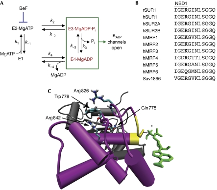
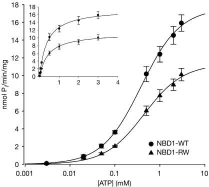
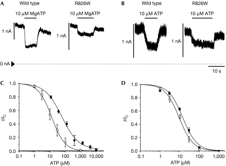
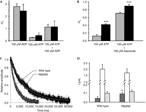
Activating mutations in the pore-forming Kir6.2 (KCNJ11) and regulatory sulphonylurea receptor SUR1 (ABCC8) subunits of the K(ATP) channel are a common cause of transient neonatal diabetes mellitus (TNDM). We identified a new TNDM mutation (R826W) in the first nucleotide-binding domain (NBD1) of SUR1. The mutation was found in a region that heterodimerizes with NBD2 to form catalytic site 2. Functional analysis showed that this mutation decreases MgATP hydrolysis by purified maltose-binding protein MBP-NBD1 fusion proteins. Inhibition of ATP hydrolysis by MgADP or BeF was not changed. The results indicate that the ATPase cycle lingers in the post-hydrolytic MgADP.P(i)-bound state, which is associated with channel activation. The extent of MgADP-dependent activation of K(ATP) channel activity was unaffected by the R826W mutation, but the time course of deactivation was slowed. Channel inhibition by MgATP was reduced, leading to an increase in resting whole-cell currents. In pancreatic beta cells, this would lead to less insulin secretion and thereby diabetes.


Conflict of interest statement

The authors declare that they have no conflict of interest.

Figures

Figure 1
Figure 1
Sequence and structure of site 2. (A) The SUR1 ATPase catalytic cycle. E is SUR1; E1, E2, … are various transitional states during ATP hydrolysis; k1, k−1, … are the rate constants for entering and leaving the different states. BeF is shown as trapping the cycle in the E2·MgATP state. (B) Signature sequence of NBD1 for various ABCC family proteins (consensus: LSxGQ/E). Residues equivalent to SUR1-Arg826 are shown in bold. (C) Side view along the plane of the membrane of a homology model of SUR1–NBD1. The catalytic core is shown in grey, the helical subdomain in purple and the signature sequence in yellow. MgADP, bound to site 2, is indicated in green. The R826–W778–R842 interaction helps to hold the helical subdomain together. In this view, the transmembrane domains (TMDs) contact NBD1 from above, indicating that Arg826 is positioned at the NBD–TMD interface. h, human; MRP, multidrug resistance-associated protein; NBD1, nucleotide-binding domain 1; r, rat; Sav1866, bacterial ABC protein.
Figure 2
Figure 2
ATPase activity is decreased by the R826W mutation. (A) ATPase activity of isolated MBP–NBD1. Wild type (WT): circles, n=7; R826W (RW): triangles, n=7. The lines are the best fit of the Michaelis–Menten equation to the mean data. MBP, maltose-binding protein; NBD1, nucleotide-binding domain 1.
Figure 3
Figure 3
ATP block of wild-type and mutant KATP channels. (A,B) Macroscopic wild-type and R826W KATP currents recorded at −60 mV in an excised patch in the (A) presence and (B) absence of Mg2+. The dashed line indicates the zero current level. Application of 10 μM ATP is indicated by the bar. (C,D) Mean relationship between (C) [MgATP] or (D) [ATP] and KATP current (I), expressed relative to that in the absence of nucleotide (Ic), for wild-type (open circles, n=5) and R826W (filled circles, n=5) channels. Membrane potential, −60 mV. In (C) and (D), the lines are the best fit of the Hill equation to the mean data: (C) wild type, IC50=17 μM, h=0.97; R826W, IC50=48 μM, h=0.82, a=0.02. (D) Wild type, IC50=9.5 μM, h=1.2; R826W, IC50 =14.5 μM, h=1.2.
Figure 4
Figure 4
Nucleotide and metabolic regulation of wild-type and mutant channels. (A) Mean KATP current recorded in the presence of 100 μM MgADP, 100 μM MgATP or 100 μM MgATP+100 μM MgADP (n=5). Current (I) is expressed relative to that in control solution (Ic). (B) Mean KATP current recorded in the presence of 100 μM MgATP or 100 μM MgATP+100 μM dizoxide (n=6). Current (I) is expressed relative to that in control solution (Ic). Scale bar is different from that in (A). (C) Rate of current deactivation following removal of 100 μM MgADP for wild-type (open circles) and R826W (filled circles, n=10) channels. (D) Mean steady-state whole-cell KATP currents evoked by a voltage step from –10 to –30 mV before (control, grey bars) and after (white bars) the application of 3 mM azide, and in the presence of 3 mM azide plus 0.5 mM tolbutamide (striped bars) for wild-type (n=10) and R826W channels (n=10). *P⩽0.05 against wild type (t-test).

Similar articles

Cited by

References

    1. Ashcroft FM (2007) The Walter B Cannon Lecture. ATP-sensitive K-channels and disease: from molecule to malady. Am J Physiol Endocrinol Metab 293: E880–E889 - PubMed
    1. Bienengraeber M et al. (2004) ABCC9 mutations identified in human dilated cardiomyopathy disrupt catalytic KATP channel gating. Nat Genet 36: 382–387 - PMC - PubMed
    1. Cheng Y, Prusoff WH (1973) Relationship between the inhibition constant (K1) and the concentration of inhibitor which causes 50 per cent inhibition (I50) of an enzymatic reaction. Biochem Pharmacol 22: 3099–3108 - PubMed
    1. Dawson RJ, Locher KP (2006) Structure of a bacterial multidrug ABC transporter. Nature 443: 156–157
    1. de Wet H, Rees M, Shimomura K, Aittoniemi J, Patch AM, Flanagan S, Ellard S, Hattersley AT, Sansom MSP, Ashcroft FM (2007a) Increased ATP-ase activity produced by mutations at R1380 in nucleotide-binding domain 2 of ABCC8 causes neonatal diabetes. Proc Natl Acad Sci USA 104: 18988–18992 - PMC - PubMed

Publication types

MeSH terms